Tolstik Elen, Osminkina Liubov A, Akimov Denis, Gongalsky Maksim B, Kudryavtsev Andrew A, Timoshenko Victor Yu, Heintzmann Rainer, Sivakov Vladimir, Popp Jürgen
Leibniz Institute of Photonic Technology, Jena 07745, Germany.
Physics Department, Lomonosov Moscow State University, Moscow 119991, Russia.
Int J Mol Sci. 2016 Sep 12;17(9):1536. doi: 10.3390/ijms17091536.
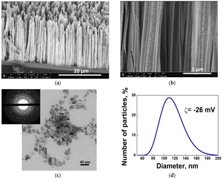
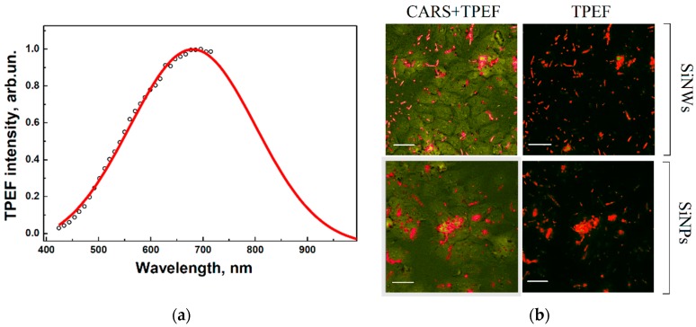
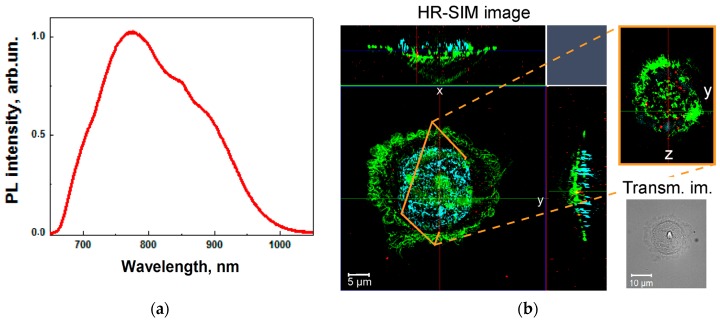
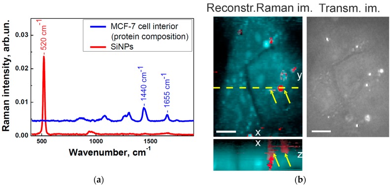
New approaches for visualisation of silicon nanoparticles (SiNPs) in cancer cells are realised by means of the linear and nonlinear optics in vitro. Aqueous colloidal solutions of SiNPs with sizes of about 10-40 nm obtained by ultrasound grinding of silicon nanowires were introduced into breast cancer cells (MCF-7 cell line). Further, the time-varying nanoparticles enclosed in cell structures were visualised by high-resolution structured illumination microscopy (HR-SIM) and micro-Raman spectroscopy. Additionally, the nonlinear optical methods of two-photon excited fluorescence (TPEF) and coherent anti-Stokes Raman scattering (CARS) with infrared laser excitation were applied to study the localisation of SiNPs in cells. Advantages of the nonlinear methods, such as rapid imaging, which prevents cells from overheating and larger penetration depth compared to the single-photon excited HR-SIM, are discussed. The obtained results reveal new perspectives of the multimodal visualisation and precise detection of the uptake of biodegradable non-toxic SiNPs by cancer cells and they are discussed in view of future applications for the optical diagnostics of cancer tumours.
通过体外线性和非线性光学手段实现了可视化癌细胞中硅纳米颗粒(SiNPs)的新方法。通过超声研磨硅纳米线获得的尺寸约为10 - 40 nm的SiNPs水胶体溶液被引入乳腺癌细胞(MCF - 7细胞系)。此外,利用高分辨率结构光照明显微镜(HR - SIM)和显微拉曼光谱对包裹在细胞结构中的随时间变化的纳米颗粒进行了可视化。另外,应用双光子激发荧光(TPEF)和红外激光激发的相干反斯托克斯拉曼散射(CARS)等非线性光学方法研究了SiNPs在细胞中的定位。讨论了非线性方法的优点,如快速成像可防止细胞过热以及与单光子激发的HR - SIM相比具有更大的穿透深度。所获得的结果揭示了多模态可视化以及精确检测癌细胞摄取可生物降解无毒SiNPs的新前景,并针对癌症肿瘤的光学诊断未来应用进行了讨论。