Cunningham Charles H, Lau Justin Y C, Chen Albert P, Geraghty Benjamin J, Perks William J, Roifman Idan, Wright Graham A, Connelly Kim A
From the Physical Sciences, Sunnybrook Research Institute, Toronto, ON, Canada (C.H.C., J.Y.C.L., B.J.G., G.A.W.); Medical Biophysics, University of Toronto, ON, Canada (C.H.C., J.Y.C.L., B.J.G., G.A.W.); GE Healthcare, Toronto, ON, Canada (A.P.C.); Pharmacy (W.J.P.) and Schulich Heart Program (I.R., G.A.W.), Sunnybrook Health Sciences Centre, Toronto, ON, Canada; and Keenan Research Centre for Biomedical Science, St. Michael's Hospital, Toronto, ON, Canada (K.A.C).
Circ Res. 2016 Nov 11;119(11):1177-1182. doi: 10.1161/CIRCRESAHA.116.309769. Epub 2016 Sep 15.
Altered cardiac energetics is known to play an important role in the progression toward heart failure. A noninvasive method for imaging metabolic markers that could be used in longitudinal studies would be useful for understanding therapeutic approaches that target metabolism.
To demonstrate the first hyperpolarized C metabolic magnetic resonance imaging of the human heart.
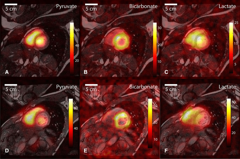
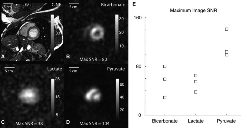
Four healthy subjects underwent conventional proton cardiac magnetic resonance imaging followed by C imaging and spectroscopic acquisition immediately after intravenous administration of a 0.1 mmol/kg dose of hyperpolarized [1-C]pyruvate. All subjects tolerated the procedure well with no adverse effects reported ≤1 month post procedure. The [1-C]pyruvate signal appeared within the chambers but not within the muscle. Imaging of the downstream metabolites showed C-bicarbonate signal mainly confined to the left ventricular myocardium, whereas the [1-C]lactate signal appeared both within the chambers and in the myocardium. The mean C image signal:noise ratio was 115 for [1-C]pyruvate, 56 for C-bicarbonate, and 53 for [1-C]lactate.
These results represent the first C images of the human heart. The appearance of C-bicarbonate signal after administration of hyperpolarized [1-C]pyruvate was readily detected in this healthy cohort (n=4). This shows that assessment of pyruvate metabolism in vivo in humans is feasible using current technology.
URL: https://www.clinicaltrials.gov. Unique identifier: NCT02648009.
已知心脏能量代谢改变在心力衰竭进展中起重要作用。一种可用于纵向研究的无创代谢标志物成像方法,对于理解针对代谢的治疗方法将很有用。
展示人类心脏的首次超极化碳代谢磁共振成像。
4名健康受试者先接受常规质子心脏磁共振成像,然后在静脉注射0.1 mmol/kg剂量的超极化[1-碳]丙酮酸后立即进行碳成像和光谱采集。所有受试者对该操作耐受性良好,术后≤1个月均未报告不良反应。[1-碳]丙酮酸信号出现在心腔内而非心肌内。下游代谢物成像显示,碳-碳酸氢盐信号主要局限于左心室心肌,而[1-碳]乳酸信号在心腔和心肌内均有出现。[1-碳]丙酮酸的平均碳图像信噪比为115,碳-碳酸氢盐为56,[1-碳]乳酸为53。
这些结果代表了人类心脏的首张碳图像。在这个健康队列(n = 4)中,超极化[1-碳]丙酮酸给药后碳-碳酸氢盐信号的出现很容易被检测到。这表明使用当前技术在人体体内评估丙酮酸代谢是可行的。