Lebbe A, Cadenas de Llano-Pérula M, Thevissen P, Verdonck A, Fieuws S, Willems G
Department of Oral Health Sciences-Orthodontics, KU Leuven and Dentistry, University Hospitals Leuven, Kapucijnenvoer 7, 3000, Leuven, Belgium.
Department of Oral Health Sciences-Forensic Dentistry, KU Leuven and Dentistry, University Hospitals Leuven, Kapucijnenvoer 7, Leuven, 3000, Belgium.
Int J Legal Med. 2017 Mar;131(2):537-546. doi: 10.1007/s00414-016-1450-0. Epub 2016 Sep 18.
Recent research concerning tooth development and dental agenesis suggests that specific genes are associated with agenesis, and that these genetic factors could also cause delayed dental development of the remaining teeth. The objective of this study was to evaluate whether dental development of patients with agenesis is delayed, compared to a control group.
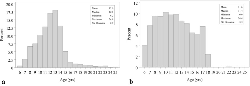
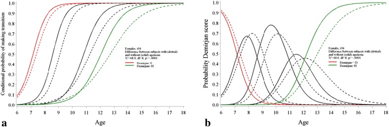
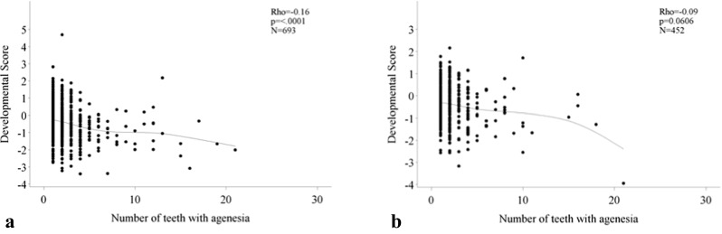
Panoramic radiographs of 1145 patients with dental agenesis were collected (452 males, 693 females) aged 6.2 to 24.8 years. The control group included 2032 panoramic radiographs (977 males, 1055 females) aged 6.0 to 24.4 years. A total of 3177 orthopantomograms were staged according to Demirjian. All left permanent teeth present in the mandible (except third molars) were considered. In order to evaluate the difference between patients with and without agenesis, a developmental score (DS) was calculated. The association between the DS and the number of agenetic teeth was evaluated with a Spearman correlation.
Based on the DS, patients with agenesis have a delayed development compared to patients in the control group (p < 0.0001). Within the agenesis group, there is a weak relation between the number of agenetic teeth and the DS: the higher the number of teeth with agenesis, the lower the DS (p < 0.0001 and p = 0.06 for females and males, respectively).
The obtained results can be an important factor for treatment planning in patients with dental agenesis. Moreover, the presence of agenesis needs to be taken into account when using age estimation methods based on permanent tooth development.
近期有关牙齿发育和牙缺失的研究表明,特定基因与牙缺失相关,且这些遗传因素也可能导致其余牙齿的发育延迟。本研究的目的是评估与对照组相比,牙缺失患者的牙齿发育是否延迟。
收集了1145例年龄在6.2至24.8岁之间的牙缺失患者的全景X线片(男性452例,女性693例)。对照组包括2032例年龄在6.0至24.4岁之间的全景X线片(男性977例,女性1055例)。根据德米尔坚法对总共3177张曲面断层片进行分期。纳入所有下颌存在的恒牙(第三磨牙除外)。为了评估有牙缺失和无牙缺失患者之间的差异,计算了发育评分(DS)。采用Spearman相关性分析评估DS与牙缺失牙齿数量之间的关联。
基于DS,与对照组患者相比,牙缺失患者的发育延迟(p < 0.0001)。在牙缺失组内,牙缺失牙齿数量与DS之间存在弱相关性:牙缺失牙齿数量越多,DS越低(女性和男性的p值分别为p < 0.0001和p = 0.06)。
所得结果可能是牙缺失患者治疗计划的一个重要因素。此外,在使用基于恒牙发育的年龄估计方法时,需要考虑牙缺失的存在。