Haridas Viraga, Ranjbar Shahin, Vorobjev Ivan A, Goldfeld Anne E, Barteneva Natasha S
Program in Cellular and Molecular Medicine, Boston Children's Hospital, Harvard Medical School, United States; Department of Pediatrics, Harvard Medical School, United States.
School of Science and Technology, Nazarbayev University, Kazakhstan; A.N. Belozersky Institute of Physico-Chemical Biology, M.V. Lomonosov Moscow State University, Russia; Department of Cell Biology and Histology, M.V. Lomonosov Moscow State University, Russia.
Methods. 2017 Jan 1;112:91-104. doi: 10.1016/j.ymeth.2016.09.007. Epub 2016 Sep 15.
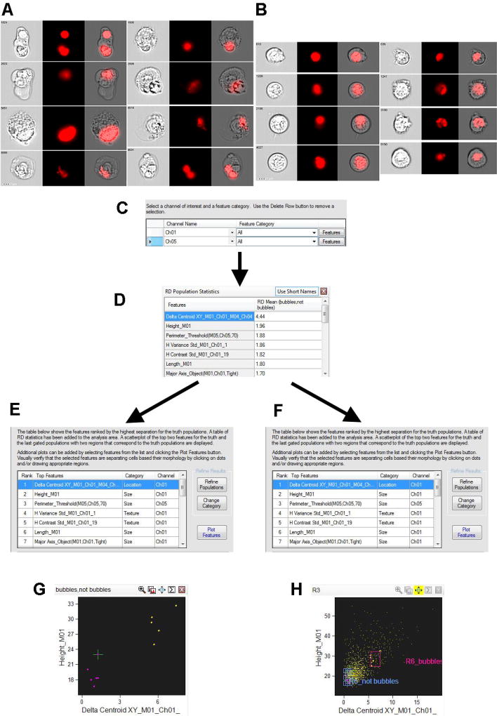
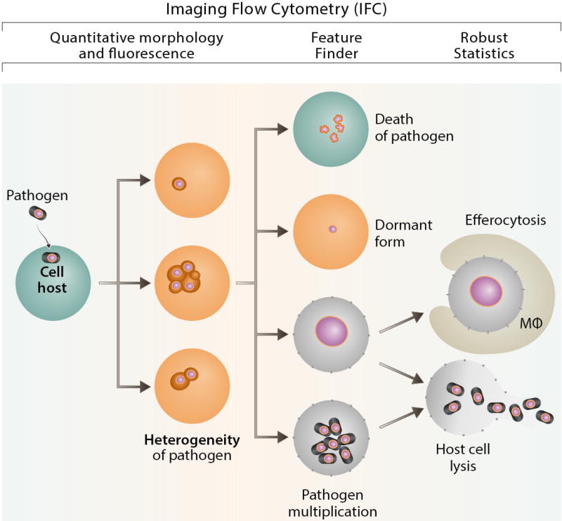
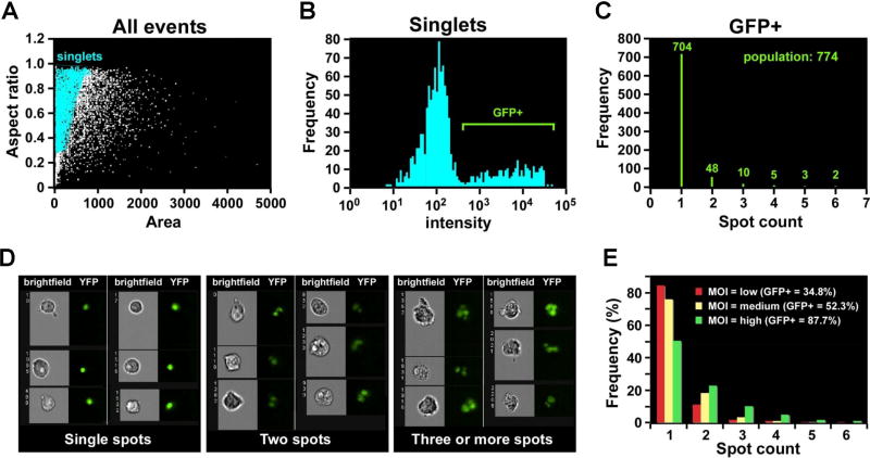
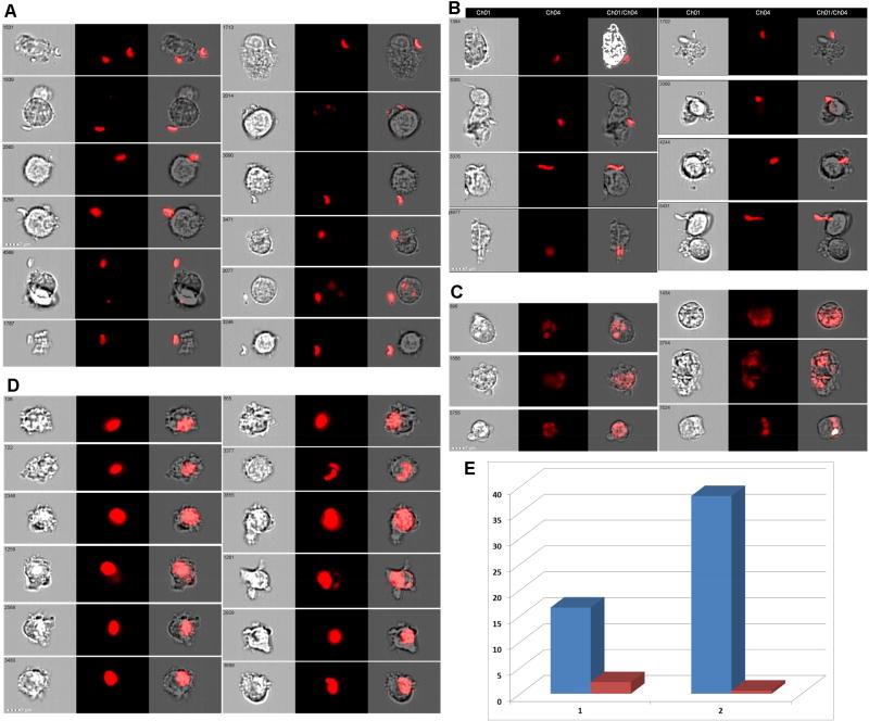
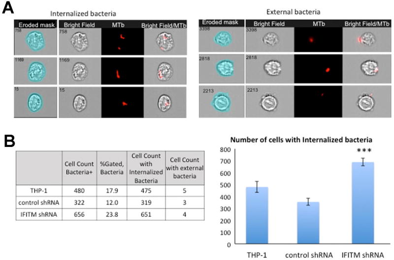
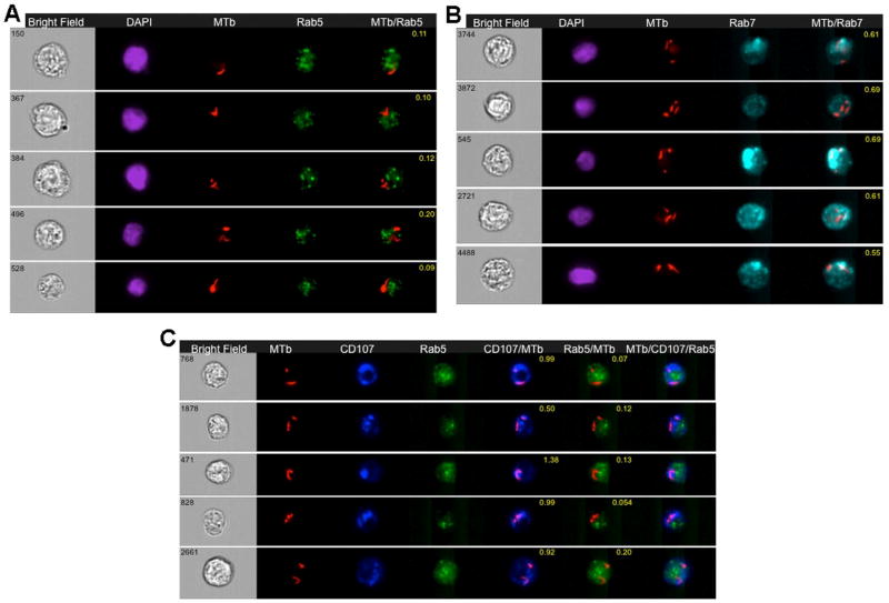
Imaging flow cytometry has been applied to address questions in infection biology, in particular, infections induced by intracellular pathogens. This methodology, which utilizes specialized analytic software makes it possible to analyze hundreds of quantified features for hundreds of thousands of individual cellular or subcellular events in a single experiment. Imaging flow cytometry analysis of host cell-pathogen interaction can thus quantitatively addresses a variety of biological questions related to intracellular infection, including cell counting, internalization score, and subcellular patterns of co-localization. Here, we provide an overview of recent achievements in the use of fluorescently labeled prokaryotic or eukaryotic pathogens in human cellular infections in analysis of host-pathogen interactions. Specifically, we give examples of Imagestream-based analysis of cell lines infected with Toxoplasma gondii or Mycobacterium tuberculosis. Furthermore, we illustrate the capabilities of imaging flow cytometry using a combination of standard IDEAS™ software and the more recently developed Feature Finder algorithm, which is capable of identifying statistically significant differences between researcher-defined image galleries. We argue that the combination of imaging flow cytometry with these software platforms provides a powerful new approach to understanding host control of intracellular pathogens.
成像流式细胞术已被应用于解决感染生物学中的问题,特别是细胞内病原体引起的感染。这种利用专门分析软件的方法,使得在单个实验中能够分析数十万个单个细胞或亚细胞事件的数百个量化特征成为可能。因此,宿主细胞与病原体相互作用的成像流式细胞术分析能够定量地解决与细胞内感染相关的各种生物学问题,包括细胞计数、内化评分和共定位的亚细胞模式。在这里,我们概述了在人类细胞感染中使用荧光标记的原核或真核病原体分析宿主-病原体相互作用的最新成果。具体来说,我们给出了基于图像流式细胞术分析感染弓形虫或结核分枝杆菌的细胞系的例子。此外,我们使用标准的IDEAS™软件和最近开发的特征查找算法的组合来说明成像流式细胞术的功能,该算法能够识别研究者定义的图像库之间的统计学显著差异。我们认为,成像流式细胞术与这些软件平台的结合为理解宿主对细胞内病原体的控制提供了一种强大的新方法。