Wang Yao, Liu Yu-Zhang, Wang Shi-Yi, Wang Zhiru
Institute and Key Laboratory of Brain Functional Genomics of Chinese Ministry of Education, Shanghai Key Laboratory of Brain Functional Genomics, School of Life Sciences, East China Normal University, Shanghai, 200062, China.
Mol Brain. 2016 Sep 29;9(1):86. doi: 10.1186/s13041-016-0266-7.
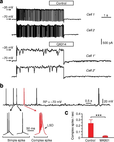
As a critical technique for dissection of synaptic and cellular mechanisms, whole-cell patch-clamp recording has become feasible for in vivo preparations including both anaesthetized and awake mammalian brains. However, compared with in vitro whole-cell recording, in vivo whole-cell recording often suffers from low success rates and high access resistance, preventing its wide application in physiological analysis of neural circuits. Here, we describe experimental procedures for achieving in vivo amphotericin B-perforated whole-cell recording as well as conventional (breakthrough) whole-cell recording from rats and mice. The success rate of perforated whole-cell recordings was 70-80 % in the hippocampus and neocortex, and access resistance was 40-70 MΩ. The success rate of conventional whole-cell recordings was ~50 % in the hippocampus, with access resistance of 20-40 MΩ. Recordings were stable, and in awake, head-fixed animals, ~50 % whole-cell patched neurons could be held for > 1 hr. The conventional whole-cell recording also permitted infusion of pharmacological agents, such as intracellular blockers of Na channels and NMDA receptors. These findings open new possibilities for synaptic and cellular analysis in vivo.
作为剖析突触和细胞机制的一项关键技术,全细胞膜片钳记录对于包括麻醉和清醒状态下的哺乳动物脑在内的体内标本而言已变得可行。然而,与体外全细胞记录相比,体内全细胞记录常常成功率较低且接入电阻较高,这阻碍了其在神经回路生理分析中的广泛应用。在此,我们描述了从大鼠和小鼠实现体内两性霉素B穿孔全细胞记录以及传统(突破式)全细胞记录的实验步骤。穿孔全细胞记录在海马体和新皮质中的成功率为70% - 80%,接入电阻为40 - 70 MΩ。传统全细胞记录在海马体中的成功率约为50%,接入电阻为20 - 40 MΩ。记录稳定,在清醒、头部固定的动物中,约50%的全细胞钳制神经元能够保持超过1小时。传统全细胞记录还允许注入药理试剂,如细胞内钠通道和NMDA受体阻滞剂。这些发现为体内突触和细胞分析开辟了新的可能性。