Jiang Jiang, Li Zhuoran, Wang Hongjun, Wang Yue, Carlson Mark A, Teusink Matthew J, MacEwan Matthew R, Gu Linxia, Xie Jingwei
Department of Surgery-Transplant and Mary & Dick Holland Regenerative Medicine Program, University of Nebraska Medical Center, Omaha, NE, 68198, USA.
Department of Mechanical and Materials Engineering, University of Nebraska-Lincoln, Lincoln, NE, 68588, USA.
Adv Healthc Mater. 2016 Dec;5(23):2993-3003. doi: 10.1002/adhm.201600808. Epub 2016 Oct 6.
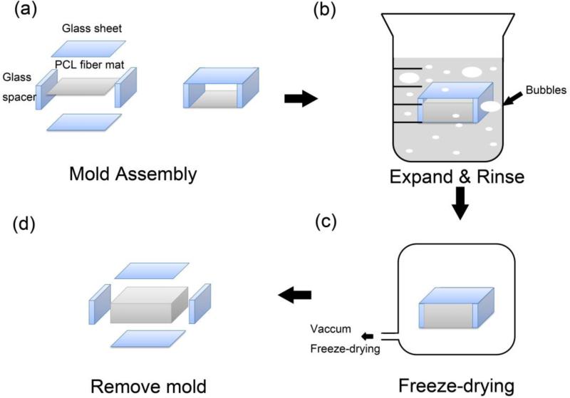
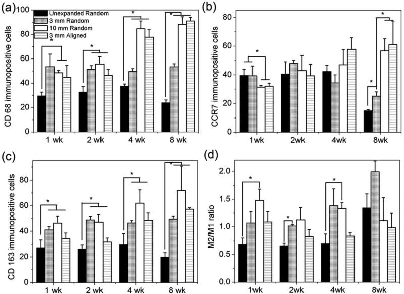
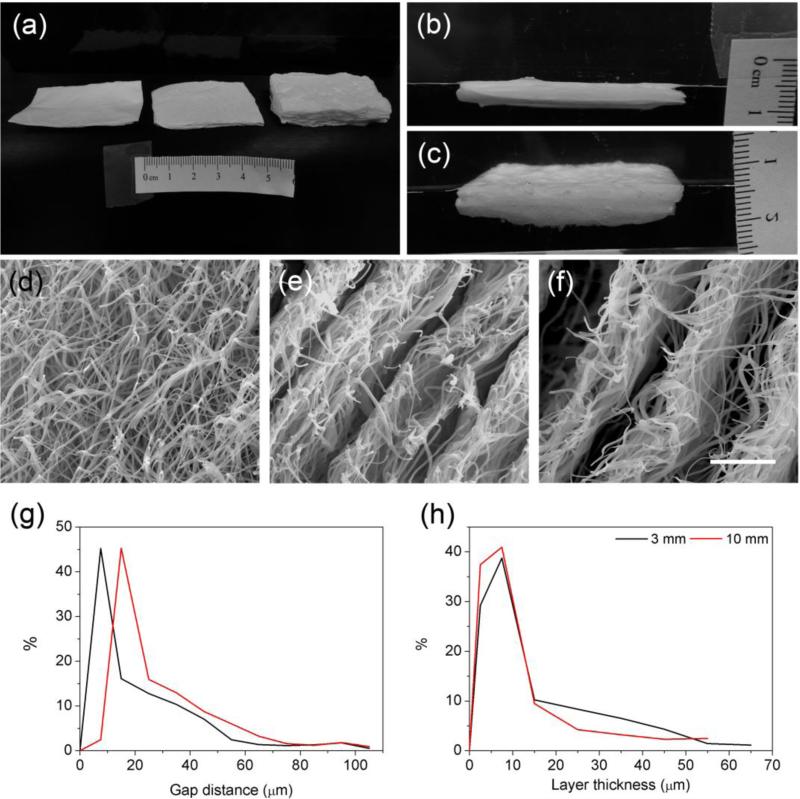
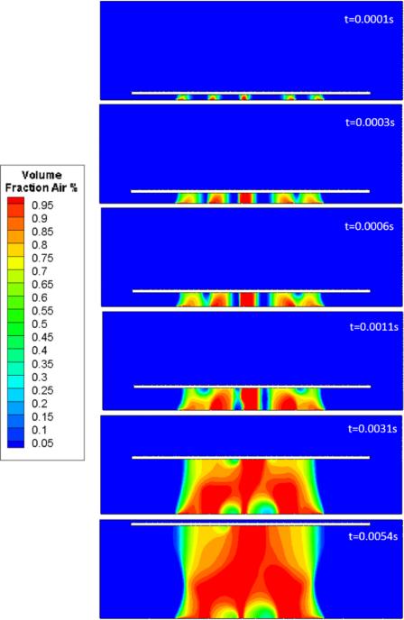
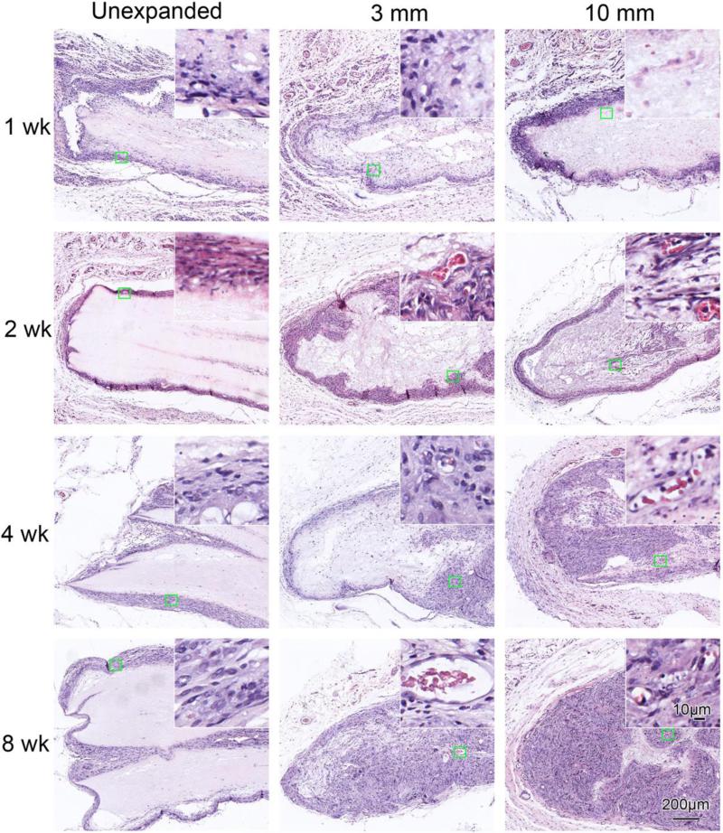
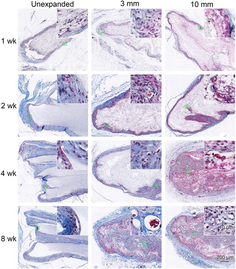
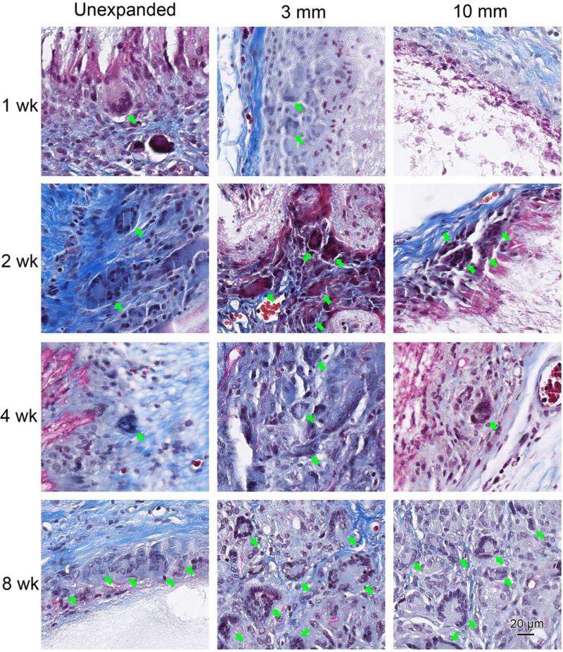
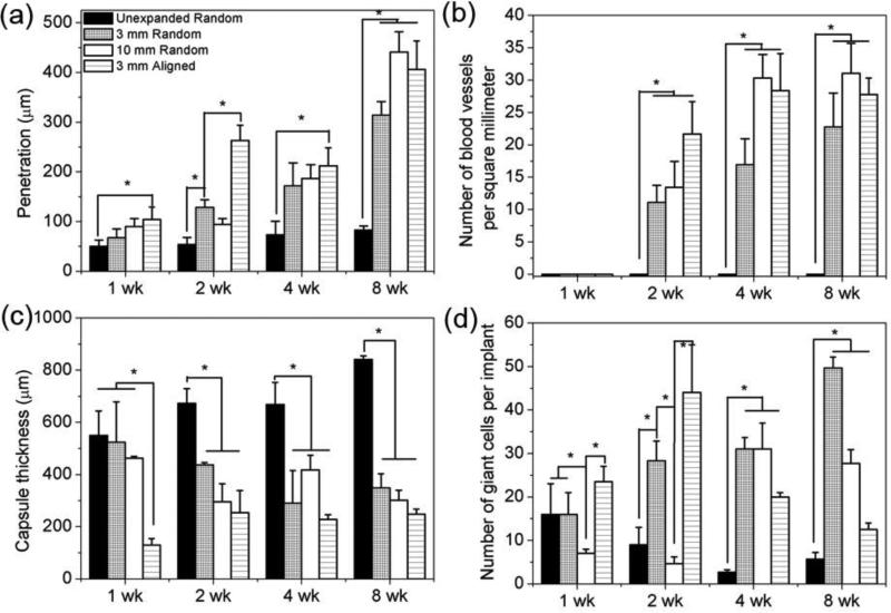
Herein, a robust method to fabricate expanded nanofiber scaffolds with controlled size and thickness using a customized mold during the modified gas-foaming process is reported. The expansion of nanofiber membranes is also simulated using a computational fluid model. Expanded nanofiber scaffolds implanted subcutaneously in rats show cellular infiltration, whereas non-expanded scaffolds only have surface cellular attachment. Compared to unexpanded nanofiber scaffolds, more CD68 and CD163 cells are observed within expanded scaffolds at all tested time points post-implantation. More CCR7 cells appear within expanded scaffolds at week 8 post-implantation. In addition, new blood vessels are present within the expanded scaffolds at week 2. The formed multinucleated giant cells within expanded scaffolds are heterogeneous expressing CD68, CCR7, or CD163 markers. Together, the present study demonstrates that the expanded nanofiber scaffolds promote cellular infiltration/tissue integration, a regenerative response, and neovascularization after subcutaneous implantation in rats. The use of expanded electrospun nanofiber scaffolds offers a promising method for in situ tissue repair/regeneration and generation of 3D tissue models/constructs.
在此,报道了一种在改进的气体发泡过程中使用定制模具制造具有可控尺寸和厚度的膨胀纳米纤维支架的稳健方法。还使用计算流体模型模拟了纳米纤维膜的膨胀。植入大鼠皮下的膨胀纳米纤维支架显示出细胞浸润,而未膨胀的支架仅具有表面细胞附着。与未膨胀的纳米纤维支架相比,在植入后的所有测试时间点,膨胀支架内观察到更多的CD68和CD163细胞。在植入后第8周,膨胀支架内出现更多的CCR7细胞。此外,在第2周时膨胀支架内存在新血管。膨胀支架内形成的多核巨细胞异质性表达CD68、CCR7或CD163标记物。总之,本研究表明,膨胀纳米纤维支架在大鼠皮下植入后促进细胞浸润/组织整合、再生反应和新血管形成。使用膨胀的电纺纳米纤维支架为原位组织修复/再生以及3D组织模型/构建体的生成提供了一种有前景的方法。