Houck Jon M, Çetin Mustafa S, Mayer Andrew R, Bustillo Juan R, Stephen Julia, Aine Cheryl, Cañive Jose, Perrone-Bizzozero Nora, Thoma Robert J, Brookes Matthew J, Calhoun Vince D
Center on Alcoholism, Substance Abuse, and Addictions, University of New Mexico, Albuquerque, New Mexico, United States; Mind Research Network, Albuquerque, New Mexico, United States.
Center on Alcoholism, Substance Abuse, and Addictions, University of New Mexico, Albuquerque, New Mexico, United States; Mind Research Network, Albuquerque, New Mexico, United States; Department of Electrical and Computer Engineering, University of New Mexico, Albuquerque, New Mexico, United States.
Neuroimage. 2017 Jan 15;145(Pt A):96-106. doi: 10.1016/j.neuroimage.2016.10.011. Epub 2016 Oct 8.
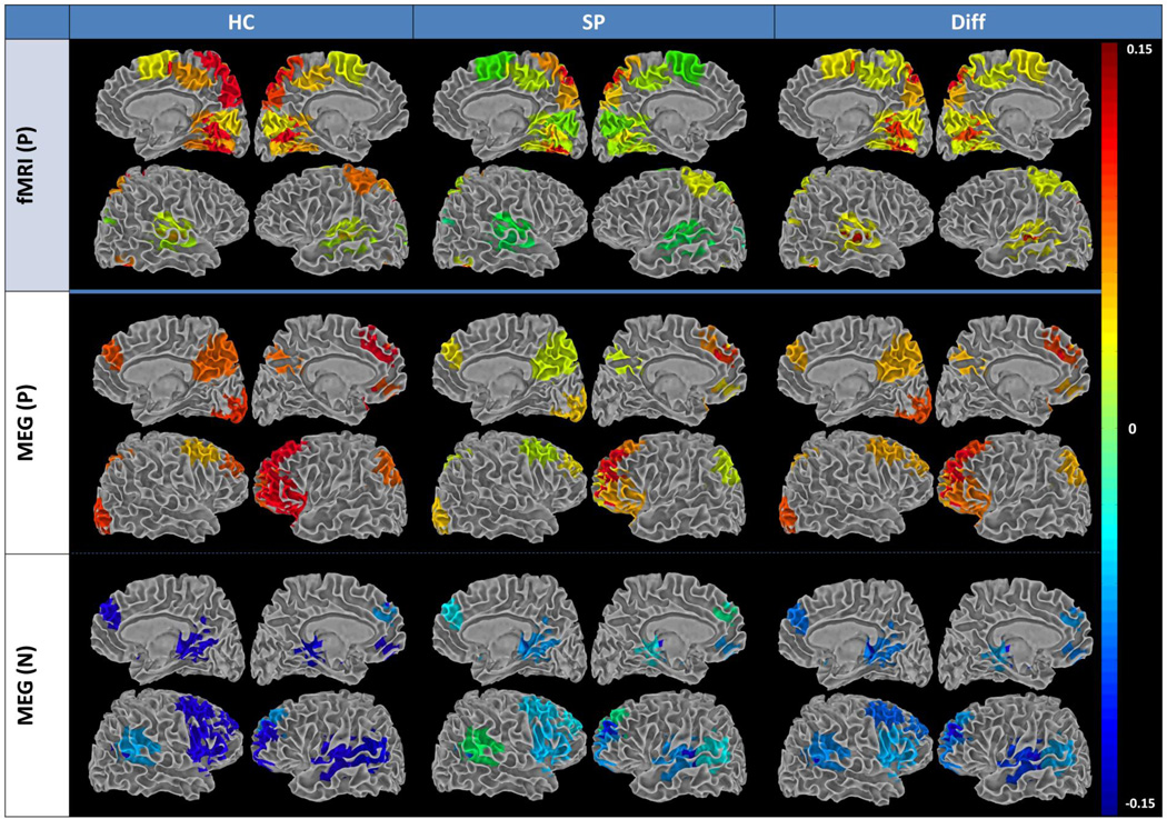
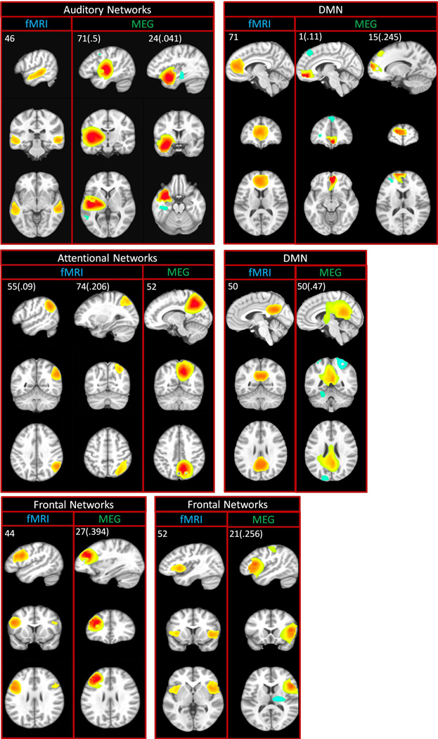
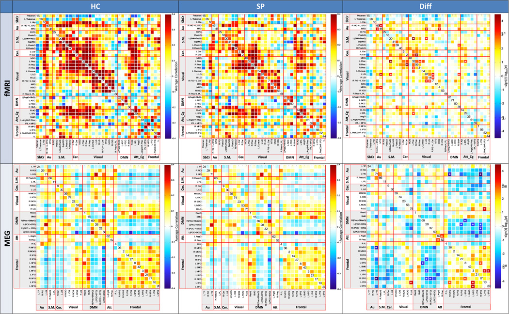
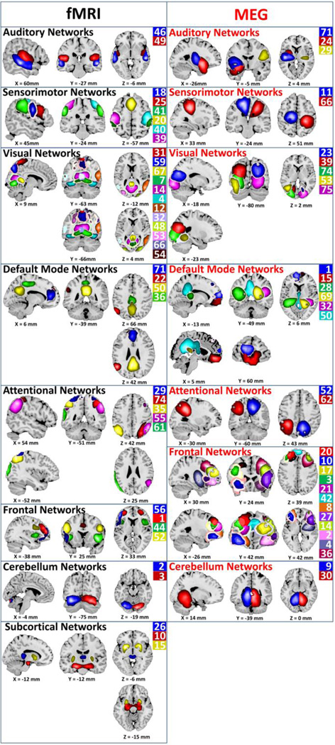
Examination of intrinsic functional connectivity using functional MRI (fMRI) has provided important findings regarding dysconnectivity in schizophrenia. Extending these results using a complementary neuroimaging modality, magnetoencephalography (MEG), we present the first direct comparison of functional connectivity between schizophrenia patients and controls, using these two modalities combined. We developed a novel MEG approach for estimation of networks using MEG that incorporates spatial independent component analysis (ICA) and pairwise correlations between independent component timecourses, to estimate intra- and intern-network connectivity. This analysis enables group-level inference and testing of between-group differences. Resting state MEG and fMRI data were acquired from a large sample of healthy controls (n=45) and schizophrenia patients (n=46). Group spatial ICA was performed on fMRI and MEG data to extract intrinsic fMRI and MEG networks and to compensate for signal leakage in MEG. Similar, but not identical spatial independent components were detected for MEG and fMRI. Analysis of functional network connectivity (FNC; i.e., pairwise correlations in network (ICA component) timecourses) revealed a differential between-modalities pattern, with greater connectivity among occipital networks in fMRI and among frontal networks in MEG. Most importantly, significant differences between controls and patients were observed in both modalities. MEG FNC results in particular indicated dysfunctional hyperconnectivity within frontal and temporal networks in patients, while in fMRI FNC was always greater for controls than for patients. This is the first study to apply group spatial ICA as an approach to leakage correction, and as such our results may be biased by spatial leakage effects. Results suggest that combining these two neuroimaging modalities reveals additional disease-relevant patterns of connectivity that were not detectable with fMRI or MEG alone.
使用功能磁共振成像(fMRI)对内在功能连接性进行检查,已得出有关精神分裂症中连接性障碍的重要发现。我们使用一种互补的神经成像方式——脑磁图(MEG)来扩展这些结果,首次对精神分裂症患者和对照组之间的功能连接性进行了直接比较,同时使用了这两种方式。我们开发了一种新颖的MEG方法来估计网络,该方法结合了空间独立成分分析(ICA)和独立成分时间序列之间的成对相关性,以估计网络内和网络间的连接性。这种分析能够进行组水平的推断并检验组间差异。静息态MEG和fMRI数据来自大量健康对照组(n = 45)和精神分裂症患者(n = 46)。对fMRI和MEG数据进行组空间ICA,以提取内在的fMRI和MEG网络,并补偿MEG中的信号泄漏。在MEG和fMRI中检测到相似但不完全相同的空间独立成分。对功能网络连接性(FNC;即网络(ICA成分)时间序列中的成对相关性)的分析揭示了一种不同模态的模式,fMRI中枕叶网络之间的连接性更强,而MEG中额叶网络之间的连接性更强。最重要的是,在两种模态中均观察到对照组和患者之间存在显著差异。特别是MEG FNC结果表明患者额叶和颞叶网络内存在功能失调的高连接性,而在fMRI中,对照组的FNC总是高于患者。这是第一项将组空间ICA作为一种泄漏校正方法应用的研究,因此我们的结果可能受到空间泄漏效应的影响。结果表明,结合这两种神经成像模态可揭示单独使用fMRI或MEG无法检测到的与疾病相关的额外连接模式。