School of Medicine, China Medical University, No. 91, Hsueh-Shih Road, Taichung, Taiwan 40402, R.O.C.
Division of Cardiovascular Medicine, Department of Medicine, China Medical University Hospital, No. 2, Yude Road, Taichung Taiwan 40447, R.O.C.
Sci Rep. 2016 Oct 17;6:35372. doi: 10.1038/srep35372.
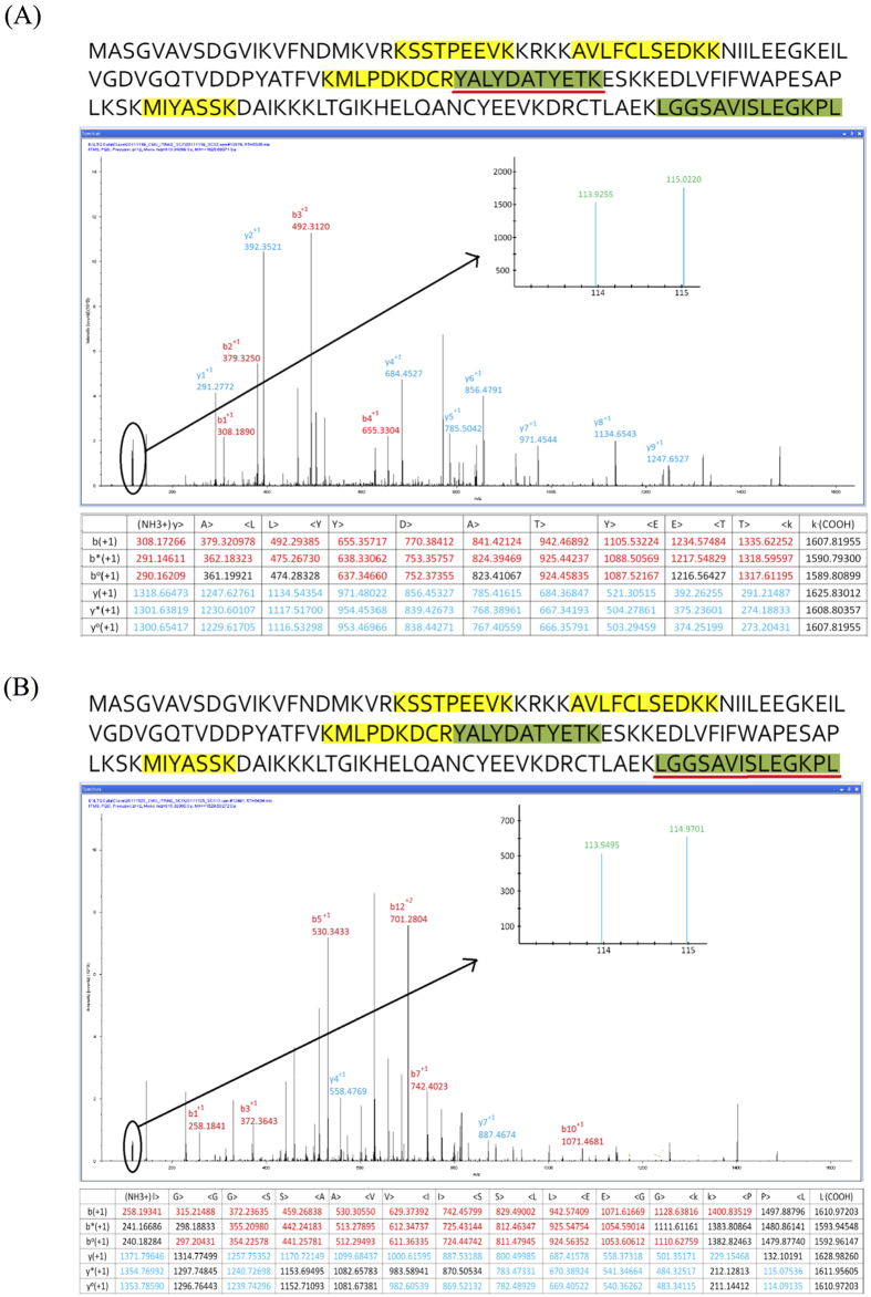
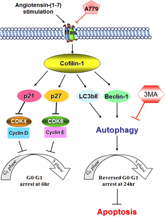
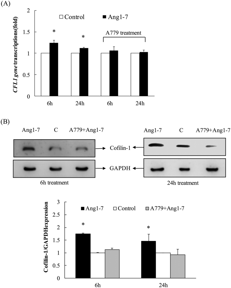
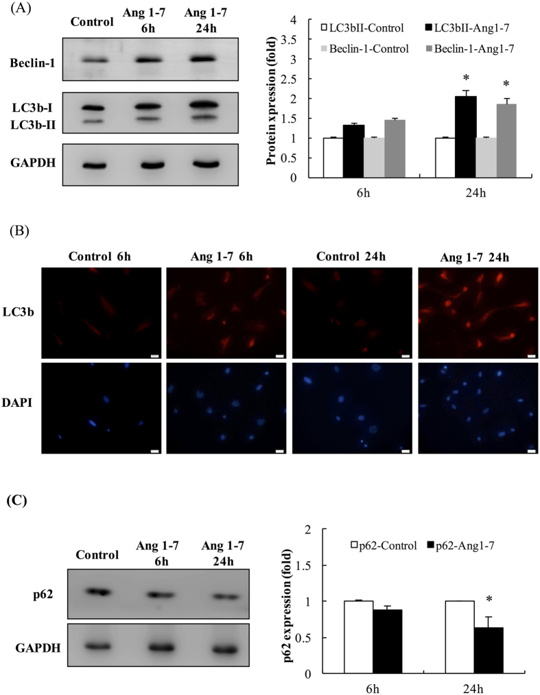
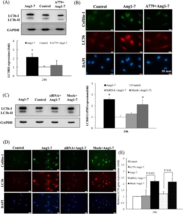
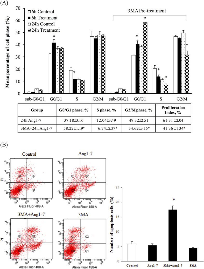
The angiotensin-converting enzyme 2/angiotensin-(1-7)/Mas axis is a pathway that acts against the detrimental effects of the renin-angiotensin system. However, the effects of angiotensin-(1-7) on endothelial protein expression and the related phenotypes are unclear. We performed a duplicate of iTRAQ quantitative proteomic analysis on human aortic endothelial cells (HAECs) treated with angiotensin-(1-7) for 6 hours. Cofilin-1 was identified as a highly abundant candidate with consistent >30% coverage and >1.2-fold overexpression in the angiotensin-(1-7)-treated group. Gene ontology analysis showed that the "regulation_of_mitosis" was significantly altered, and cell cycle analysis indicated that the 6-hour angiotensin-(1-7) treatment significantly induced G0/G1 arrest. Knockdown of the cofilin-1 (CFL1) gene suggested the G0/G1 phase arrest was mediated by the modulation of p27 and the p21/Cyclin/CDK complex by Cofilin-1. Interestingly, quiescent HAECs escaped G0/G1 arrest upon angiotensin-(1-7) treatment for 24 hours, and angiotensin-(1-7) induced autophagy by upregulating Beclin-1 and microtubule-associated protein 1 light chain 3b-II expression, which was also attenuated by A779 pre-treatment and CFL1 knockdown. After pre-treatment with 3-methyladenine (3MA), treatment with angiotensin-(1-7) for 24 h induced significant G0/G1 phase arrest and apoptosis, suggesting a pro-survival role of autophagy in this context. In conclusion, Cofilin-1 plays a dominant role in angiotensin-(1-7)-induced G0/G1 arrest and autophagy to maintain cellular homeostasis in HAECs.
血管紧张素转换酶 2/血管紧张素-(1-7)/Mas 轴是一种对抗肾素-血管紧张素系统有害作用的途径。然而,血管紧张素-(1-7)对内皮蛋白表达和相关表型的影响尚不清楚。我们对用血管紧张素-(1-7)处理 6 小时的人主动脉内皮细胞(HAEC)进行了两次 iTRAQ 定量蛋白质组学分析。原肌球蛋白-1被鉴定为一个高度丰富的候选物,在血管紧张素-(1-7)处理组中的覆盖率>30%,表达水平上调>1.2 倍。基因本体分析显示,“有丝分裂调控”显著改变,细胞周期分析表明,6 小时血管紧张素-(1-7)处理显著诱导 G0/G1 期阻滞。原肌球蛋白-1(CFL1)基因的敲低表明,G0/G1 期阻滞是由 Cofilin-1 对 p27 和 p21/Cyclin/CDK 复合物的调节介导的。有趣的是,静息的 HAEC 在血管紧张素-(1-7)处理 24 小时后逃脱了 G0/G1 期阻滞,血管紧张素-(1-7)通过上调 Beclin-1 和微管相关蛋白 1 轻链 3b-II 的表达诱导自噬,这一过程也被 A779 预处理和 CFL1 敲低所减弱。在用 3-甲基腺嘌呤(3MA)预处理后,用血管紧张素-(1-7)处理 24 小时会诱导明显的 G0/G1 期阻滞和细胞凋亡,这表明自噬在这种情况下具有促进存活的作用。总之,Cofilin-1 在血管紧张素-(1-7)诱导的 G0/G1 期阻滞和自噬中发挥主导作用,以维持 HAEC 中的细胞内稳态。