Zhong Xin, Aredo Bogale, Ding Yi, Zhang Kaiyan, Zhao Cynthia X, Ufret-Vincenty Rafael L
Department of Ophthalmology, University of Texas Southwestern Medical Center, Dallas, Texas, United States.
Invest Ophthalmol Vis Sci. 2016 Oct 1;57(13):5558-5567. doi: 10.1167/iovs.16-19965.
Oxidative stress, partly due to light, has an important role in many retinal diseases, including macular degeneration and retinal dystrophies. The Leu450Met variant of RPE65 is expressed in C57BL/6 and in many genetically modified mice. It confers significant resistance to light induced retinal degeneration (LIRD). Our goal was to develop an effective and efficient method to induce LIRD in resistant mice that would recapitulate mechanisms seen in known models of LIRD.
The retinas of C57BL/6J mice were exposed to light using a murine fundus camera. Two protocols (with and without intraperitoneal fluorescein) were used. Optical coherence tomography (OCT) helped determine the location and extent of retinal damage. Histology, TUNEL assay, quantitative (q) PCR, and immunohistochemistry were performed.
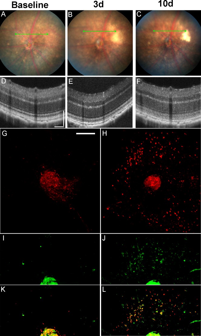
Both protocols consistently generated LIRD in C57BL/6J mice. Optical coherence tomography and histology demonstrated that retinal damage starts at the level of the photoreceptor/outer retina and is more prominent in the superior retina. Fundus camera-delivered light-induced retinal degeneration (FCD-LIRD) is associated with apoptosis, subretinal microglia/macrophages, increased expression of oxidative stress response genes, and C3d deposition.
We characterize two new models of light-induced retinal degeneration that are effective in C57BL/6J mice, and can be modulated in terms of severity. We expect FCD-LIRD to be useful in exploring mechanisms of LIRD in resistant mice, which will be important in increasing our understanding of the retinal response to light damage and oxidative stress.
氧化应激在包括黄斑变性和视网膜营养不良在内的许多视网膜疾病中起重要作用,部分原因是光照。RPE65的Leu450Met变体在C57BL/6和许多基因工程小鼠中表达。它赋予对光诱导视网膜变性(LIRD)的显著抗性。我们的目标是开发一种有效且高效的方法,在抗性小鼠中诱导LIRD,该方法将重现已知LIRD模型中所见的机制。
使用鼠眼底相机对C57BL/6J小鼠的视网膜进行光照。采用了两种方案(有和没有腹腔注射荧光素)。光学相干断层扫描(OCT)有助于确定视网膜损伤的位置和程度。进行了组织学、TUNEL检测、定量(q)PCR和免疫组织化学分析。
两种方案均在C57BL/6J小鼠中持续产生LIRD。光学相干断层扫描和组织学表明,视网膜损伤始于光感受器/外视网膜水平,且在视网膜上半部分更明显。眼底相机介导的光诱导视网膜变性(FCD-LIRD)与细胞凋亡、视网膜下小胶质细胞/巨噬细胞、氧化应激反应基因表达增加以及C3d沉积有关。
我们描述了两种在C57BL/6J小鼠中有效的光诱导视网膜变性新模型,并且可以在严重程度方面进行调节。我们期望FCD-LIRD在探索抗性小鼠中LIRD的机制方面有用,这对于增进我们对视网膜对光损伤和氧化应激反应的理解将很重要。