Tremblay Yannick D N, Labrie Josée, Chénier Sonia, Jacques Mario
Groupe de recherche sur les maladies infectieuses du porc, Faculté de médecine vétérinaire, Université de Montréal, St-Hyacinthe, QC, J2S 2M2, Canada.
Laboratoire d'épidémiosurveillance animale du Québec, Ministère de l'Agriculture, des Pêcheries et de l'Alimentation du Québec, St-Hyacinthe, QC, J2S 7X9, Canada.
Microb Biotechnol. 2017 Jul;10(4):756-760. doi: 10.1111/1751-7915.12432. Epub 2016 Oct 28.
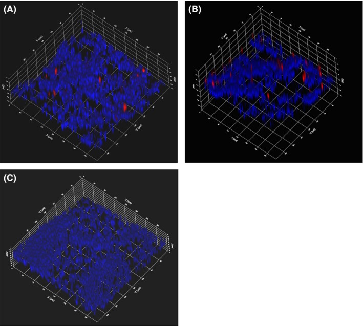
Actinobacillus pleuropneumoniae causes porcine pleuropneumonia and forms biofilms in vitro on abiotic surfaces; however, presence of biofilms during infections has not been documented. The aim of this study was to use a species-specific fluorescent oligonucleotide probe and confocal microscopy to localize A. pleuropneumoniae in the lungs of two naturally infected pigs. Actinobacillus pleuropneumoniae was detected by fluorescence in situ hybridization and observed to grow as aggregates (~30-45 μm) during a natural infection. As the A. pleuropneumoniae aggregates observed in porcine lungs differed from the biofilms grown on a solid surface obtained in vitro, we designed a new biofilm assay using agarose, a porous substrate, favouring the formation of aggregates. In this study, we described for the first time the mode of growth of A. pleuropneumoniae during a natural infection in pigs. We also propose an in vitro biofilm assay for A. pleuropneumoniae using a porous substrate which allows the formation of aggregates. This assay might be more representative of the in vivo situation, at least in terms of the size of the bacterial aggregates and the presence of a porous matrix, and could potentially be used to test the susceptibility of A. pleuropneumoniae aggregates to antibiotics and disinfectants.
胸膜肺炎放线杆菌可引发猪胸膜肺炎,并能在体外非生物表面形成生物膜;然而,感染过程中生物膜的存在尚未见文献记载。本研究的目的是使用种特异性荧光寡核苷酸探针和共聚焦显微镜,在两只自然感染猪的肺中定位胸膜肺炎放线杆菌。通过荧光原位杂交检测到胸膜肺炎放线杆菌,并观察到其在自然感染期间以聚集体形式(约30 - 45μm)生长。由于在猪肺中观察到的胸膜肺炎放线杆菌聚集体与体外在固体表面生长的生物膜不同,我们设计了一种新的生物膜检测方法,使用琼脂糖这种多孔基质,有利于聚集体的形成。在本研究中,我们首次描述了胸膜肺炎放线杆菌在猪自然感染期间的生长方式。我们还提出了一种针对胸膜肺炎放线杆菌的体外生物膜检测方法,使用多孔基质,可形成聚集体。该检测方法可能更能代表体内情况,至少在细菌聚集体大小和多孔基质存在方面,并且有可能用于测试胸膜肺炎放线杆菌聚集体对抗生素和消毒剂的敏感性。