• 文献检索
  • 文档翻译
  • 深度研究
  • 学术资讯
  • Suppr Zotero 插件Zotero 插件
  • 邀请有礼
  • 套餐&价格
  • 历史记录
应用&插件
Suppr Zotero 插件Zotero 插件浏览器插件Mac 客户端Windows 客户端微信小程序
定价
高级版会员购买积分包购买API积分包
服务
文献检索文档翻译深度研究API 文档MCP 服务
关于我们
关于 Suppr公司介绍联系我们用户协议隐私条款
关注我们

Suppr 超能文献

核心技术专利:CN118964589B侵权必究
粤ICP备2023148730 号-1Suppr @ 2026

文献检索

告别复杂PubMed语法,用中文像聊天一样搜索,搜遍4000万医学文献。AI智能推荐,让科研检索更轻松。

立即免费搜索

文件翻译

保留排版,准确专业,支持PDF/Word/PPT等文件格式,支持 12+语言互译。

免费翻译文档

深度研究

AI帮你快速写综述,25分钟生成高质量综述,智能提取关键信息,辅助科研写作。

立即免费体验

通过生物发光成像和荧光断层扫描对脑肿瘤生长进行体内随访

In Vivo Follow-up of Brain Tumor Growth via Bioluminescence Imaging and Fluorescence Tomography.

作者信息

Genevois Coralie, Loiseau Hugues, Couillaud Franck

机构信息

Molecular Imaging and Innovative Therapy in Oncology (IMOTION), EA 7435, University of Bordeaux, Bordeaux 33076, France.

出版信息

Int J Mol Sci. 2016 Oct 31;17(11):1815. doi: 10.3390/ijms17111815.

DOI:10.3390/ijms17111815
PMID:27809256
原文链接:https://pmc.ncbi.nlm.nih.gov/articles/PMC5133816/
Abstract

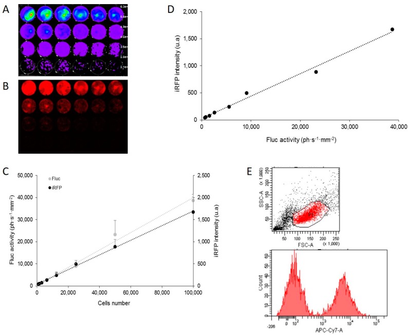
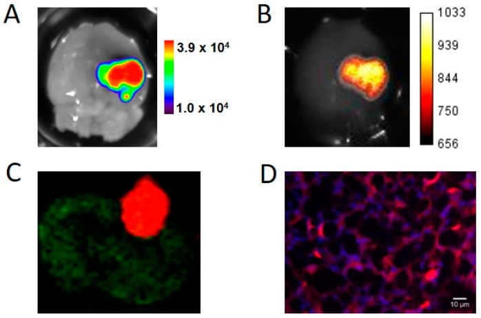
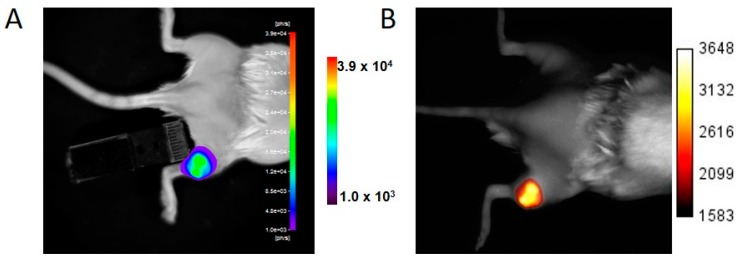
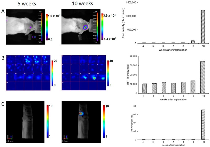
Reporter gene-based strategies are widely used in experimental oncology. Bioluminescence imaging (BLI) using the firefly luciferase (Fluc) as a reporter gene and d-luciferin as a substrate is currently the most widely employed technique. The present paper compares the performances of BLI imaging with fluorescence imaging using the near infrared fluorescent protein (iRFP) to monitor brain tumor growth in mice. Fluorescence imaging includes fluorescence reflectance imaging (FRI), fluorescence diffuse tomography (fDOT), and fluorescence molecular Imaging (FMT). A U87 cell line was genetically modified for constitutive expression of both the encoding Fluc and iRFP reporter genes and assayed for cell, subcutaneous tumor and brain tumor imaging. On cultured cells, BLI was more sensitive than FRI; in vivo, tumors were first detected by BLI. Fluorescence of iRFP provided convenient tools such as flux cytometry, direct detection of the fluorescent protein on histological slices, and fluorescent tomography that allowed for 3D localization and absolute quantification of the fluorescent signal in brain tumors.

摘要

基于报告基因的策略在实验肿瘤学中被广泛应用。使用萤火虫荧光素酶(Fluc)作为报告基因、D - 荧光素作为底物的生物发光成像(BLI)是目前应用最广泛的技术。本文比较了BLI成像与使用近红外荧光蛋白(iRFP)的荧光成像在监测小鼠脑肿瘤生长方面的性能。荧光成像包括荧光反射成像(FRI)、荧光扩散断层扫描(fDOT)和荧光分子成像(FMT)。对U87细胞系进行基因改造,使其组成性表达编码Fluc和iRFP的报告基因,并用于细胞、皮下肿瘤和脑肿瘤成像检测。在培养细胞上,BLI比FRI更敏感;在体内,肿瘤首先通过BLI被检测到。iRFP的荧光提供了便捷工具,如流式细胞术、在组织切片上直接检测荧光蛋白以及荧光断层扫描,后者可实现脑肿瘤中荧光信号的三维定位和绝对定量。

https://cdn.ncbi.nlm.nih.gov/pmc/blobs/dc44/5133816/e7d36e5310e6/ijms-17-01815-g004.jpg
https://cdn.ncbi.nlm.nih.gov/pmc/blobs/dc44/5133816/58eee599208e/ijms-17-01815-g001.jpg
https://cdn.ncbi.nlm.nih.gov/pmc/blobs/dc44/5133816/5eaee578f0f8/ijms-17-01815-g002a.jpg
https://cdn.ncbi.nlm.nih.gov/pmc/blobs/dc44/5133816/dfe1a8a45ccf/ijms-17-01815-g003.jpg
https://cdn.ncbi.nlm.nih.gov/pmc/blobs/dc44/5133816/e7d36e5310e6/ijms-17-01815-g004.jpg

相似文献

1
In Vivo Follow-up of Brain Tumor Growth via Bioluminescence Imaging and Fluorescence Tomography.通过生物发光成像和荧光断层扫描对脑肿瘤生长进行体内随访
Int J Mol Sci. 2016 Oct 31;17(11):1815. doi: 10.3390/ijms17111815.
2
Detection of Brain Tumors and Systemic Metastases Using NanoLuc and Fluc for Dual Reporter Imaging.使用纳米荧光素酶和萤火虫荧光素酶进行双报告基因成像检测脑肿瘤和全身转移灶
Mol Imaging Biol. 2016 Feb;18(1):62-9. doi: 10.1007/s11307-015-0864-2.
3
Noninvasive bioluminescence imaging of luciferase expressing intracranial U87 xenografts: correlation with magnetic resonance imaging determined tumor volume and longitudinal use in assessing tumor growth and antiangiogenic treatment effect.表达荧光素酶的颅内U87异种移植瘤的无创生物发光成像:与磁共振成像测定的肿瘤体积的相关性以及在评估肿瘤生长和抗血管生成治疗效果中的纵向应用
Neurosurgery. 2006 Feb;58(2):365-72; discussion 365-72. doi: 10.1227/01.NEU.0000195114.24819.4F.
4
Fluorescence and Bioluminescence Imaging of Orthotopic Brain Tumors in Mice.小鼠原位脑肿瘤的荧光和生物发光成像
Methods Mol Biol. 2017;1530:283-305. doi: 10.1007/978-1-4939-6646-2_17.
5
Optimized Longitudinal Monitoring of Stem Cell Grafts in Mouse Brain Using a Novel Bioluminescent/Near Infrared Fluorescent Fusion Reporter.优化利用新型生物发光/近红外荧光融合报告基因对小鼠脑内干细胞移植物的纵向监测。
Cell Transplant. 2017 Dec;26(12):1878-1889. doi: 10.1177/0963689717739718.
6
Detection of brain tumors using fluorescence diffuse optical tomography and nanoparticles as contrast agents.使用荧光漫射光学断层成像术和纳米颗粒作为对比剂检测脑瘤。
J Biomed Opt. 2012 Dec;17(12):126004. doi: 10.1117/1.JBO.17.12.126004.
7
In vivo bioluminescence imaging.体内生物发光成像。
Comp Med. 2004 Dec;54(6):631-4.
8
Near-infrared bioluminescent proteins for two-color multimodal imaging.用于双色多模态成像的近红外生物发光蛋白。
Sci Rep. 2016 Nov 11;6:36588. doi: 10.1038/srep36588.
9
Evaluating the effects of immunosuppression by in-vivo bioluminescence imaging after allotransplantation of ovarian grafts.移植卵巢移植物后体内生物发光成像评估免疫抑制作用。
Reprod Biomed Online. 2011 Feb;22(2):220-7. doi: 10.1016/j.rbmo.2010.10.010. Epub 2010 Oct 31.
10
A luciferin analogue generating near-infrared bioluminescence achieves highly sensitive deep-tissue imaging.一种产生近红外生物发光的荧光素类似物可实现高灵敏度的深层组织成像。
Nat Commun. 2016 Jun 14;7:11856. doi: 10.1038/ncomms11856.

引用本文的文献

1
Development of an optimized machine learning approach for assessing brain metastatic burden in preclinical models.开发一种用于评估临床前模型中脑转移负担的优化机器学习方法。
bioRxiv. 2024 Aug 23:2024.08.21.608131. doi: 10.1101/2024.08.21.608131.
2
Inhibition of orthotopic castration-resistant prostate cancer growth and metastasis in mice by JC VLPs carrying a suicide gene driven by the PSA promoter.载有 PSA 启动子驱动的自杀基因的 JC VLPs 抑制小鼠原位去势抵抗性前列腺癌生长和转移。
Cancer Gene Ther. 2024 Feb;31(2):250-258. doi: 10.1038/s41417-023-00699-8. Epub 2023 Dec 11.
3
Long-term in vivo chimeric cells tracking in non-human primate.

本文引用的文献

1
Using Dual Fluorescence Reporting Genes to Establish an In Vivo Imaging Model of Orthotopic Lung Adenocarcinoma in Mice.利用双荧光报告基因建立小鼠原位肺腺癌体内成像模型。
Mol Imaging Biol. 2016 Dec;18(6):849-859. doi: 10.1007/s11307-016-0967-4.
2
Optical imaging probes in oncology.肿瘤学中的光学成像探针。
Oncotarget. 2016 Jul 26;7(30):48753-48787. doi: 10.18632/oncotarget.9066.
3
Detection of Brain Tumors and Systemic Metastases Using NanoLuc and Fluc for Dual Reporter Imaging.使用纳米荧光素酶和萤火虫荧光素酶进行双报告基因成像检测脑肿瘤和全身转移灶
在非人灵长类动物中长期体内嵌合细胞的示踪。
Protein Cell. 2024 Feb 29;15(3):207-222. doi: 10.1093/procel/pwad049.
4
Imaging Techniques in Pharmacological Precision Medicine.药理学精准医学中的影像学技术。
Handb Exp Pharmacol. 2023;280:213-235. doi: 10.1007/164_2023_641.
5
New insights into detecting alizarin from autofluorescence in marked glass eels.从标记玻璃鳗的自发荧光中检测茜素的新见解。
Sci Rep. 2022 Sep 26;12(1):15985. doi: 10.1038/s41598-022-18440-0.
6
Factors Influencing Luciferase-Based Bioluminescent Imaging in Preclinical Models of Brain Tumor.影响脑肿瘤临床前模型中基于荧光素酶的生物发光成像的因素。
Drug Metab Dispos. 2022 Mar;50(3):277-286. doi: 10.1124/dmd.121.000597. Epub 2021 Dec 9.
7
Indocyanine Green-Camptothecin Co-Loaded Perfluorocarbon Double-Layer Nanocomposite: A Versatile Nanotheranostics for Photochemotherapy and FDOT Diagnosis of Breast Cancer.吲哚菁绿-喜树碱共负载全氟碳双层纳米复合材料:一种用于乳腺癌光化学疗法和FDOT诊断的多功能纳米诊疗剂
Pharmaceutics. 2021 Sep 17;13(9):1499. doi: 10.3390/pharmaceutics13091499.
8
iRFP (near-infrared fluorescent protein) imaging of subcutaneous and deep tissue tumours in mice highlights differences between imaging platforms.对小鼠皮下和深部组织肿瘤进行的红外荧光蛋白(iRFP)成像突出了成像平台之间的差异。
Cancer Cell Int. 2021 May 3;21(1):247. doi: 10.1186/s12935-021-01918-8.
9
Novel NanoLuc substrates enable bright two-population bioluminescence imaging in animals.新型 NanoLuc 底物可实现动物体内明亮的双种群生物发光成像。
Nat Methods. 2020 Aug;17(8):852-860. doi: 10.1038/s41592-020-0889-6. Epub 2020 Jul 13.
10
Kinetic modeling and analysis of dynamic bioluminescence imaging of substrates administered by intraperitoneal injection.腹腔注射给药底物的动态生物发光成像的动力学建模与分析
Quant Imaging Med Surg. 2020 Feb;10(2):389-396. doi: 10.21037/qims.2020.01.01.
Mol Imaging Biol. 2016 Feb;18(1):62-9. doi: 10.1007/s11307-015-0864-2.
4
Noninvasive near-infrared fluorescent protein-based imaging of tumor progression and metastases in deep organs and intraosseous tissues.基于非侵入性近红外荧光蛋白的深部器官和骨内组织肿瘤进展及转移成像。
J Biomed Opt. 2014 Jan;19(1):16019. doi: 10.1117/1.JBO.19.1.016019.
5
iRFP is a sensitive marker for cell number and tumor growth in high-throughput systems.红外荧光蛋白(iRFP)是高通量系统中细胞数量和肿瘤生长的敏感标志物。
Cell Cycle. 2014;13(2):220-6. doi: 10.4161/cc.26985. Epub 2013 Oct 31.
6
In vivo imaging of orthotopic prostate cancer with far-red gene reporter fluorescence tomography and in vivo and ex vivo validation.利用远红基因报告荧光体层成像术对原位前列腺癌进行体内成像及体内和体外验证。
J Biomed Opt. 2013 Oct;18(10):101305. doi: 10.1117/1.JBO.18.10.101305.
7
Near-infrared fluorescent proteins for multicolor in vivo imaging.近红外荧光蛋白用于活体多色成像。
Nat Methods. 2013 Aug;10(8):751-4. doi: 10.1038/nmeth.2521. Epub 2013 Jun 16.
8
Tumor margin detection using quantitative NIRF molecular imaging targeting EpCAM validated by far red gene reporter iRFP.使用针对 EpCAM 的定量近红外荧光分子成像进行肿瘤边缘检测,远红基因报告 iRFP 验证。
Mol Imaging Biol. 2013 Oct;15(5):560-8. doi: 10.1007/s11307-013-0637-8.
9
Detection of brain tumors using fluorescence diffuse optical tomography and nanoparticles as contrast agents.使用荧光漫射光学断层成像术和纳米颗粒作为对比剂检测脑瘤。
J Biomed Opt. 2012 Dec;17(12):126004. doi: 10.1117/1.JBO.17.12.126004.
10
Bright and stable near-infrared fluorescent protein for in vivo imaging.用于活体成像的明亮且稳定的近红外荧光蛋白。
Nat Biotechnol. 2011 Jul 17;29(8):757-61. doi: 10.1038/nbt.1918.