Yao Amy, Pain Margaret, Balchandani Priti, Shrivastava Raj K
Department of Neurosurgery, Icahn School of Medicine at Mount Sinai, Annenberg 8, One Gustave L Levy Pl, New York, NY, 10029, USA.
Neurosurg Rev. 2018 Jul;41(3):745-753. doi: 10.1007/s10143-016-0801-0. Epub 2016 Nov 21.
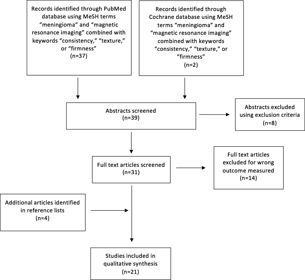
Tumor consistency is a critical factor that influences operative strategy and patient counseling. Magnetic resonance imaging (MRI) describes the concentration of water within living tissues and as such, is hypothesized to predict aspects of their biomechanical behavior. In meningiomas, MRI signal intensity has been used to predict the consistency of the tumor and its histopathological subtype, though its predictive capacity is debated in the literature. We performed a systematic review of the PubMed database since 1990 concerning MRI appearance and tumor consistency to assess whether or not MRI can be used reliably to predict tumor firmness. The inclusion criteria were case series and clinical studies that described attempts to correlate preoperative MRI findings with tumor consistency. The relationship between the pre-operative imaging characteristics, intraoperative findings, and World Health Organization (WHO) histopathological subtype is described. While T2 signal intensity and MR elastography provide a useful predictive measure of tumor consistency, other techniques have not been validated. T1-weighted imaging was not found to offer any diagnostic or predictive value. A quantitative assessment of T2 signal intensity more reliably predicts consistency than inherently variable qualitative analyses. Preoperative knowledge of tumor firmness affords the neurosurgeon substantial benefit when planning surgical techniques. Based upon our review of the literature, we currently recommend the use of T2-weighted MRI for predicting consistency, which has been shown to correlate well with analysis of tumor histological subtype. Development of standard measures of tumor consistency, standard MRI quantification metrics, and further exploration of MRI technique may improve the predictive ability of neuroimaging for meningiomas.
肿瘤质地是影响手术策略和患者咨询的关键因素。磁共振成像(MRI)描述了活体组织内的水浓度,因此,据推测它可以预测组织生物力学行为的各个方面。在脑膜瘤中,MRI信号强度已被用于预测肿瘤的质地及其组织病理学亚型,尽管其预测能力在文献中存在争议。我们对1990年以来的PubMed数据库进行了系统综述,内容涉及MRI表现与肿瘤质地,以评估MRI是否可可靠地用于预测肿瘤硬度。纳入标准为描述术前MRI结果与肿瘤质地相关性的病例系列和临床研究。描述了术前影像学特征、术中发现与世界卫生组织(WHO)组织病理学亚型之间的关系。虽然T2信号强度和磁共振弹性成像提供了有用的肿瘤质地预测指标,但其他技术尚未得到验证。未发现T1加权成像具有任何诊断或预测价值。与固有可变的定性分析相比,对T2信号强度进行定量评估能更可靠地预测质地。术前了解肿瘤硬度在规划手术技术时能为神经外科医生带来很大益处。基于我们对文献的综述,我们目前推荐使用T2加权MRI来预测质地,它已被证明与肿瘤组织学亚型分析有很好的相关性。制定肿瘤质地的标准测量方法、标准MRI定量指标以及进一步探索MRI技术可能会提高神经影像学对脑膜瘤的预测能力。