• 文献检索
  • 文档翻译
  • 深度研究
  • 学术资讯
  • Suppr Zotero 插件Zotero 插件
  • 邀请有礼
  • 套餐&价格
  • 历史记录
应用&插件
Suppr Zotero 插件Zotero 插件浏览器插件Mac 客户端Windows 客户端微信小程序
定价
高级版会员购买积分包购买API积分包
服务
文献检索文档翻译深度研究API 文档MCP 服务
关于我们
关于 Suppr公司介绍联系我们用户协议隐私条款
关注我们

Suppr 超能文献

核心技术专利:CN118964589B侵权必究
粤ICP备2023148730 号-1Suppr @ 2026

文献检索

告别复杂PubMed语法,用中文像聊天一样搜索,搜遍4000万医学文献。AI智能推荐,让科研检索更轻松。

立即免费搜索

文件翻译

保留排版,准确专业,支持PDF/Word/PPT等文件格式,支持 12+语言互译。

免费翻译文档

深度研究

AI帮你快速写综述,25分钟生成高质量综述,智能提取关键信息,辅助科研写作。

立即免费体验

使用增加数量的光学相干断层扫描图像测量眼前节结构的益处:华裔美国人眼研究

Benefit of Measuring Anterior Segment Structures Using an Increased Number of Optical Coherence Tomography Images: The Chinese American Eye Study.

作者信息

Xu Benjamin Y, Israelsen Paul, Pan Billy X, Wang Dandan, Jiang Xuejuan, Varma Rohit

机构信息

Roski Eye Institute, Keck School of Medicine at the University of Southern California Los Angeles, Los Angeles, California, United States.

Keck School of Medicine at the University of Southern California Los Angeles, Los Angeles, California, United States.

出版信息

Invest Ophthalmol Vis Sci. 2016 Nov 1;57(14):6313-6319. doi: 10.1167/iovs.16-19755.

DOI:10.1167/iovs.16-19755
PMID:27893097
原文链接:https://pmc.ncbi.nlm.nih.gov/articles/PMC5119487/
Abstract

PURPOSE

The purpose of this study was to evaluate the benefit of analyzing an increased number of anterior segment optical coherence tomography (AS-OCT) images on measurement values of various anterior segment parameters.

METHODS

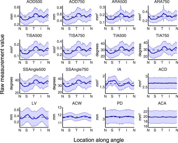
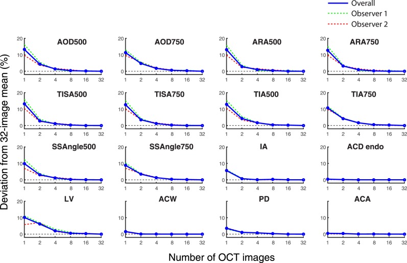
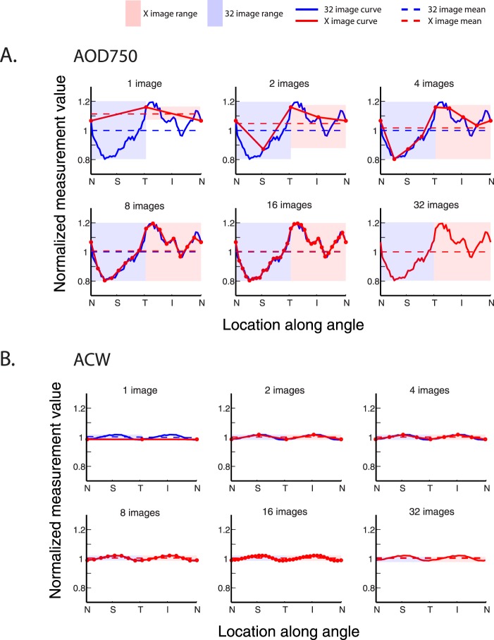
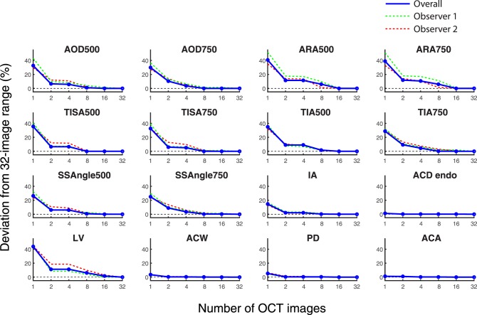
Subjects for this cross-sectional study were recruited from the Chinese American Eye Study (CHES), a population-based study in Los Angeles, CA. Thirty-two AS-OCT images were acquired from one eye each of 83 consecutive subjects. Sixteen parameters were analyzed in each image, including angle opening distance (AOD), angle recess area (ARA), trabecular iris space area (TISA), trabecular iris angle (TIA), scleral spur angle (SSAngle), lens vault (LV), pupillary diameter (PD), anterior chamber depth (ACD), anterior chamber width (ACW), iris area (IA), and anterior chamber area (ACA). Data from 1, 2, 4, 8, 16, or 32 OCT images were averaged across subjects to calculate the range and mean of measurement values for each parameter.

RESULTS

Anatomical variations were poorly captured with fewer OCT images for AOD, ARA, TISA, SSAngle, IA, and LV. For these parameters, the range and mean of measurement values obtained from one OCT image deviated from 32-image values by up to 43.9% and 13.3% of the 32-image mean, respectively. These deviations decreased when additional OCT images were analyzed. Deviations from 32-image range and mean values were less pronounced regardless of image number for PD, ACD, ACW, and ACA, measuring up to 3.5% and 5.0%, respectively.

CONCLUSIONS

A multi-image approach should be the standard in OCT-based studies of AOD, ARA, TISA, TIA, SSAngle, IA, and LV.

摘要

目的

本研究旨在评估分析更多数量的眼前节光学相干断层扫描(AS-OCT)图像对各种眼前节参数测量值的益处。

方法

本横断面研究的受试者来自加利福尼亚州洛杉矶的基于人群的华裔美国人眼研究(CHES)。连续83名受试者的每只眼睛均采集32张AS-OCT图像。每张图像分析16个参数,包括房角开放距离(AOD)、房角隐窝面积(ARA)、小梁虹膜间隙面积(TISA)、小梁虹膜角(TIA)、巩膜突角(SSAngle)、晶状体拱高(LV)、瞳孔直径(PD)、前房深度(ACD)、前房宽度(ACW)、虹膜面积(IA)和前房面积(ACA)。对1、2、4、8、16或32张OCT图像的数据进行受试者间平均,以计算每个参数测量值的范围和平均值。

结果

对于AOD、ARA、TISA、SSAngle、IA和LV,较少的OCT图像难以捕捉解剖变异。对于这些参数,从一张OCT图像获得的测量值范围和平均值分别比32张图像的值偏离32张图像平均值的43.9%和13.3%。分析额外的OCT图像时,这些偏差会减小。对于PD、ACD、ACW和ACA,无论图像数量如何,与32张图像范围和平均值的偏差均不太明显,分别高达3.5%和5.0%。

结论

在基于OCT的AOD、ARA、TISA、TIA、SSAngle、IA和LV研究中,多图像方法应成为标准。

https://cdn.ncbi.nlm.nih.gov/pmc/blobs/1901/5119487/5a6af253bc5a/i1552-5783-57-14-6313-f04.jpg
https://cdn.ncbi.nlm.nih.gov/pmc/blobs/1901/5119487/64ea90a9dda7/i1552-5783-57-14-6313-f01.jpg
https://cdn.ncbi.nlm.nih.gov/pmc/blobs/1901/5119487/c321081555da/i1552-5783-57-14-6313-f02.jpg
https://cdn.ncbi.nlm.nih.gov/pmc/blobs/1901/5119487/897a26610d49/i1552-5783-57-14-6313-f03.jpg
https://cdn.ncbi.nlm.nih.gov/pmc/blobs/1901/5119487/5a6af253bc5a/i1552-5783-57-14-6313-f04.jpg

相似文献

1
Benefit of Measuring Anterior Segment Structures Using an Increased Number of Optical Coherence Tomography Images: The Chinese American Eye Study.使用增加数量的光学相干断层扫描图像测量眼前节结构的益处:华裔美国人眼研究
Invest Ophthalmol Vis Sci. 2016 Nov 1;57(14):6313-6319. doi: 10.1167/iovs.16-19755.
2
Effect of Angle Narrowing on Sectoral Variation of Anterior Chamber Angle Width: The Chinese American Eye Study.角度变窄对前房角宽度扇形变化的影响:中美眼研究。
Ophthalmol Glaucoma. 2020 Mar-Apr;3(2):130-138. doi: 10.1016/j.ogla.2019.12.005. Epub 2019 Dec 27.
3
Repeatability and Reproducibility of Anterior Chamber Angle Measurement with Swept-Source Optical Coherence Tomography in Patients with Primary Angle Closure Suspect.在原发性闭角型青光眼疑似患者中,应用扫频源光学相干断层扫描仪测量前房角的可重复性和再现性。
Curr Eye Res. 2021 Dec;46(12):1853-1860. doi: 10.1080/02713683.2021.1942069. Epub 2021 Jul 12.
4
Analyzing Anatomical Factors Contributing to Angle Closure Based on Anterior Segment Optical Coherence Tomography Imaging.基于眼前节光学相干断层扫描成像分析导致房角关闭的解剖学因素。
Curr Eye Res. 2022 Feb;47(2):256-261. doi: 10.1080/02713683.2021.1978098. Epub 2021 Sep 27.
5
Correlation between Intraocular Pressure and Angle Configuration Measured by OCT: The Chinese American Eye Study.OCT 测量的眼压与角度结构的相关性:中美眼研究。
Ophthalmol Glaucoma. 2018 Nov-Dec;1(3):158-166. doi: 10.1016/j.ogla.2018.09.001. Epub 2018 Sep 29.
6
Effects of lens extraction versus laser peripheral iridotomy on anterior segment morphology in primary angle closure suspect.晶状体摘除术与激光周边虹膜切开术对原发性闭角型青光眼可疑患者眼前节形态的影响
Graefes Arch Clin Exp Ophthalmol. 2019 Jul;257(7):1473-1480. doi: 10.1007/s00417-019-04353-8. Epub 2019 May 11.
7
Comparison of Anterior Segment Optical Coherence Tomography Parameters Among Vietnamese, Chinese, and Whites.越南人、中国人和白种人眼前节光学相干断层扫描参数的比较。
Am J Ophthalmol. 2018 Nov;195:72-82. doi: 10.1016/j.ajo.2018.07.034. Epub 2018 Aug 4.
8
[Detection of the anterior segment configuration of primary angle-closure glaucoma by anterior segment optical coherence tomography].[眼前节光学相干断层扫描对原发性闭角型青光眼眼前节形态的检测]
Zhonghua Yan Ke Za Zhi. 2013 Feb;49(2):109-15.
9
Optimal number and orientation of anterior segment OCT images to measure ocular biometric parameters in angle closure eyes: the Chinese American Eye Study.在闭角型眼中测量眼球生物测量参数的前节 OCT 图像的最佳数量和方向:中美眼研究。
Br J Ophthalmol. 2023 Jun;107(6):795-801. doi: 10.1136/bjophthalmol-2021-319275. Epub 2022 Jan 21.
10
Diagnostic Abilities of Three-Dimensional Anterior Segment Optical Coherence Tomography in Detecting Angle Closure.三维眼前节光学相干断层扫描在检测房角关闭中的诊断能力。
J Glaucoma. 2024 Oct 1;33(10):801-807. doi: 10.1097/IJG.0000000000002442. Epub 2024 May 24.

引用本文的文献

1
Morphologic features of crystalline lens in cataract patients with different lens sclerosis and axial length.不同晶状体硬化程度和眼轴长度的白内障患者晶状体的形态学特征
BMC Ophthalmol. 2025 Jul 3;25(1):391. doi: 10.1186/s12886-025-04148-y.
2
Deep learning-based normative database of anterior chamber dimensions for angle closure assessment: the Singapore Chinese Eye Study.基于深度学习的用于房角关闭评估的前房深度规范数据库:新加坡华人眼研究
Br J Ophthalmol. 2025 Mar 20;109(4):497-503. doi: 10.1136/bjo-2024-325602.
3
Classification of Angle Closure Severity by Hierarchical Cluster Analysis of Ocular Biometrics in the Dark and Light.

本文引用的文献

1
Comparison of Physiologic versus Pharmacologic Mydriasis on Anterior Chamber Angle Measurements Using Spectral Domain Optical Coherence Tomography.使用光谱域光学相干断层扫描技术比较生理性散瞳与药物性散瞳对前房角测量的影响
J Ophthalmol. 2015;2015:845643. doi: 10.1155/2015/845643. Epub 2015 Mar 23.
2
Optimal number of angle images for calculating anterior angle volume and iris volume measurements.用于计算前房角容积和虹膜容积测量的最佳角度图像数量。
Invest Ophthalmol Vis Sci. 2015 May;56(5):2842-7. doi: 10.1167/iovs.14-15883.
3
Evaluation of filtering blebs exhibiting transconjunctival oozing using anterior segment optical coherence tomography.
在黑暗和光亮环境下的眼部生物测量的层次聚类分析对闭角型青光眼严重程度的分类
Transl Vis Sci Technol. 2023 Sep 1;12(9):4. doi: 10.1167/tvst.12.9.4.
4
Early Assessment of Circumferential Anterior Segment Structures Following Implantable Collamer Lens V4c Implantation Via SS-OCT.基于 SS-OCT 的 V4c 可折叠人工晶状体植入术后眼前节周向结构的早期评估。
Transl Vis Sci Technol. 2022 Nov 1;11(11):4. doi: 10.1167/tvst.11.11.4.
5
Biometric indicators of anterior segment parameters before and after laser peripheral iridotomy by swept-source optical coherent tomography.应用扫频源光学相干断层扫描测量激光周边虹膜切开术前后眼前节参数的生物测量指标。
BMC Ophthalmol. 2022 May 16;22(1):222. doi: 10.1186/s12886-022-02448-1.
6
Optimal number and orientation of anterior segment OCT images to measure ocular biometric parameters in angle closure eyes: the Chinese American Eye Study.在闭角型眼中测量眼球生物测量参数的前节 OCT 图像的最佳数量和方向:中美眼研究。
Br J Ophthalmol. 2023 Jun;107(6):795-801. doi: 10.1136/bjophthalmol-2021-319275. Epub 2022 Jan 21.
7
Ocular Biometric Determinants of Dark-to-Light Change in Angle Width: The Chinese American Eye Study.眼生物测量学指标对角度宽度明暗变化的影响:美国华裔眼研究。
Am J Ophthalmol. 2022 May;237:183-192. doi: 10.1016/j.ajo.2021.10.027. Epub 2021 Nov 2.
8
Ocular Biometric Risk Factors for Progression of Primary Angle Closure Disease: The Zhongshan Angle Closure Prevention Trial.原发性闭角型青光眼进展的眼生物测量危险因素:中山闭角型青光眼防治研究。
Ophthalmology. 2022 Mar;129(3):267-275. doi: 10.1016/j.ophtha.2021.10.003. Epub 2021 Oct 8.
9
Measurement of Iris Thickness at Different Regions in Healthy Chinese Adults.健康中国成年人不同区域虹膜厚度的测量
J Ophthalmol. 2021 May 11;2021:2653564. doi: 10.1155/2021/2653564. eCollection 2021.
10
Anatomic Changes and Predictors of Angle Widening after Laser Peripheral Iridotomy: The Zhongshan Angle Closure Prevention Trial.激光周边虹膜切开术后房角增宽的解剖学变化及预测因素:中山闭角防治试验。
Ophthalmology. 2021 Aug;128(8):1161-1168. doi: 10.1016/j.ophtha.2021.01.021. Epub 2021 Jan 23.
使用眼前节光学相干断层扫描技术评估出现经结膜渗液的滤过泡。
Graefes Arch Clin Exp Ophthalmol. 2015 Mar;253(3):439-45. doi: 10.1007/s00417-014-2872-3. Epub 2014 Dec 9.
4
Anterior segment optical coherence tomography parameters in phacomorphic angle closure and mature cataracts.晶状体膨胀性房角关闭和成熟白内障的眼前节光学相干断层扫描参数
Invest Ophthalmol Vis Sci. 2014 Oct 21;55(11):7403-9. doi: 10.1167/iovs.14-14748.
5
Reproducibility of anterior segment angle metrics measurements derived from Cirrus spectral domain optical coherence tomography.基于Cirrus光谱域光学相干断层扫描技术的眼前节角度测量指标的可重复性
J Glaucoma. 2015 Jun-Jul;24(5):e47-51. doi: 10.1097/IJG.0000000000000136.
6
Outcomes of laser peripheral iridotomy in angle closure subgroups according to anterior segment optical coherence tomography parameters.根据眼前节光学相干断层扫描参数,激光周边虹膜切开术在闭角型亚组中的治疗效果。
Invest Ophthalmol Vis Sci. 2014 Sep 23;55(10):6795-801. doi: 10.1167/iovs.14-14714.
7
Reproducibility of angle metrics using the time-domain anterior segment optical coherence tomography: intra-observer and inter-observer variability.使用时域眼前节光学相干断层扫描技术测量角度指标的可重复性:观察者内和观察者间的变异性。
Curr Eye Res. 2015 May;40(5):496-500. doi: 10.3109/02713683.2014.930155. Epub 2014 Jun 23.
8
Anterior Chamber Angle Assessment by Anterior-segment Optical Coherence Tomography After Phacoemulsification With or Without Goniosynechialysis in Patients With Primary Angle Closure Glaucoma.原发性闭角型青光眼患者白内障超声乳化吸除术联合或不联合房角粘连分离术后前房角的眼前节光学相干断层扫描评估
J Glaucoma. 2015 Dec;24(9):647-55. doi: 10.1097/IJG.0000000000000061.
9
Longitudinal changes of angle configuration in primary angle-closure suspects: the Zhongshan Angle-Closure Prevention Trial.原发性闭角型青光眼可疑者房角形态的纵向变化:中山闭角型青光眼预防试验
Ophthalmology. 2014 Sep;121(9):1699-1705. doi: 10.1016/j.ophtha.2014.03.039. Epub 2014 May 15.
10
Sectoral variations of iridocorneal angle width and iris volume in Chinese Singaporeans: a swept-source optical coherence tomography study.新加坡华裔人群中虹膜角膜角宽度和虹膜体积的扇形差异:一项扫频光学相干断层扫描研究
Graefes Arch Clin Exp Ophthalmol. 2014 Jul;252(7):1127-32. doi: 10.1007/s00417-014-2636-0. Epub 2014 Apr 30.