Dong Zachary M, Wollstein Gadi, Wang Bo, Schuman Joel S
University of Pittsburgh Medical Center (UPMC) Eye Center, Eye and Ear Institute, Department of Ophthalmology, University of Pittsburgh School of Medicine, Ophthalmology and Visual Science Research Center, Pittsburgh, PA, United States.
New York University (NYU) Langone Eye Center, NYU Langone Medical Center, Department of Ophthalmology, NYU School of Medicine, New York, NY, United States.
Prog Retin Eye Res. 2017 Mar;57:76-88. doi: 10.1016/j.preteyeres.2016.11.001. Epub 2016 Dec 1.
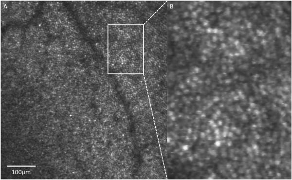
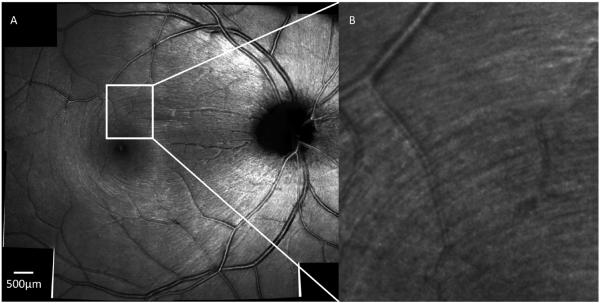
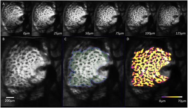
Since the introduction of commercial optical coherence tomography (OCT) systems, the ophthalmic imaging modality has rapidly expanded and it has since changed the paradigm of visualization of the retina and revolutionized the management and diagnosis of neuro-retinal diseases, including glaucoma. OCT remains a dynamic and evolving imaging modality, growing from time-domain OCT to the improved spectral-domain OCT, adapting novel image analysis and processing methods, and onto the newer swept-source OCT and the implementation of adaptive optics (AO) into OCT. The incorporation of AO into ophthalmic imaging modalities has enhanced OCT by improving image resolution and quality, particularly in the posterior segment of the eye. Although OCT previously captured in-vivo cross-sectional images with unparalleled high resolution in the axial direction, monochromatic aberrations of the eye limit transverse or lateral resolution to about 15-20 μm and reduce overall image quality. In pairing AO technology with OCT, it is now possible to obtain diffraction-limited resolution images of the optic nerve head and retina in three-dimensions, increasing resolution down to a theoretical 3 μm. It is now possible to visualize discrete structures within the posterior eye, such as photoreceptors, retinal nerve fiber layer bundles, the lamina cribrosa, and other structures relevant to glaucoma. Despite its limitations and barriers to widespread commercialization, the expanding role of AO in OCT is propelling this technology into clinical trials and onto becoming an invaluable modality in the clinician's arsenal.
自从商业光学相干断层扫描(OCT)系统问世以来,这种眼科成像方式迅速发展,改变了视网膜可视化的模式,并彻底革新了包括青光眼在内的神经视网膜疾病的管理和诊断方法。OCT仍然是一种动态发展的成像方式,从时域OCT发展到改进后的频域OCT,采用了新颖的图像分析和处理方法,进而发展到更新的扫频源OCT以及将自适应光学(AO)应用于OCT。将AO纳入眼科成像方式通过提高图像分辨率和质量增强了OCT,特别是在眼后段。尽管OCT以前能够在轴向方向以无与伦比的高分辨率获取体内横截面图像,但眼睛的单色像差将横向分辨率限制在约15 - 20微米,并降低了整体图像质量。通过将AO技术与OCT相结合,现在可以获得三维的视神经乳头和视网膜的衍射极限分辨率图像,将分辨率提高到理论上的3微米。现在可以可视化眼后段内的离散结构,如光感受器、视网膜神经纤维层束、筛板以及其他与青光眼相关的结构。尽管AO在OCT中的应用存在局限性和广泛商业化的障碍,但AO在OCT中不断扩大的作用正推动这项技术进入临床试验,并成为临床医生不可或缺的一种方式。