Centre de Résonance Magnétique des Systèmes Biologiques, UMR5536, CNRS/Université de Bordeaux, 146 rue Léo Saignat, 33076 Bordeaux, France.
Sci Rep. 2016 Dec 20;6:39449. doi: 10.1038/srep39449.
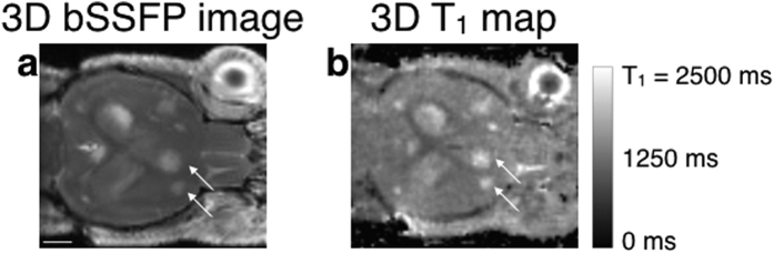
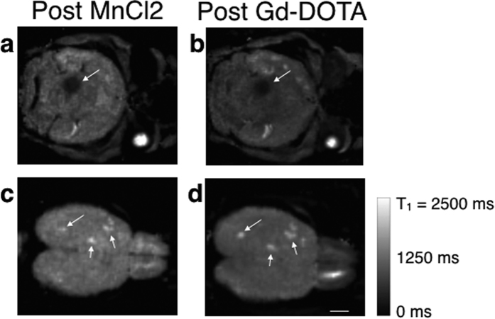
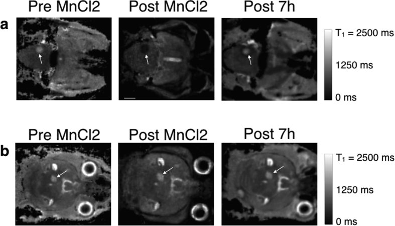
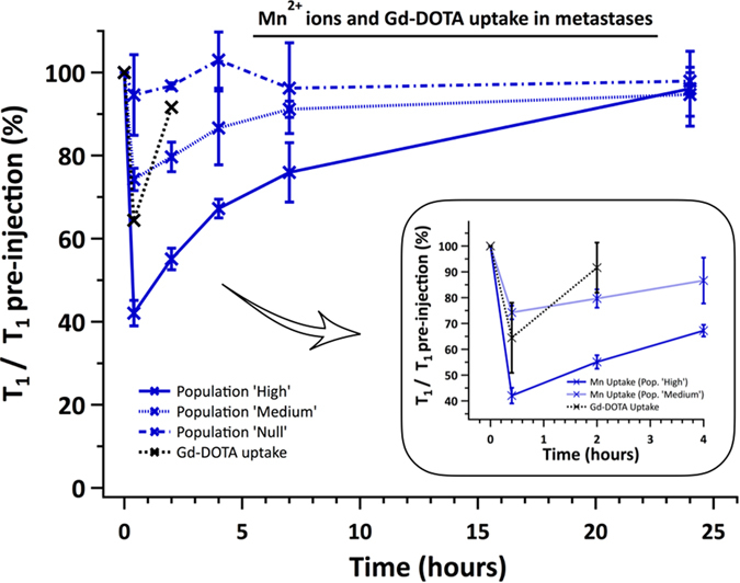
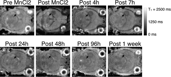
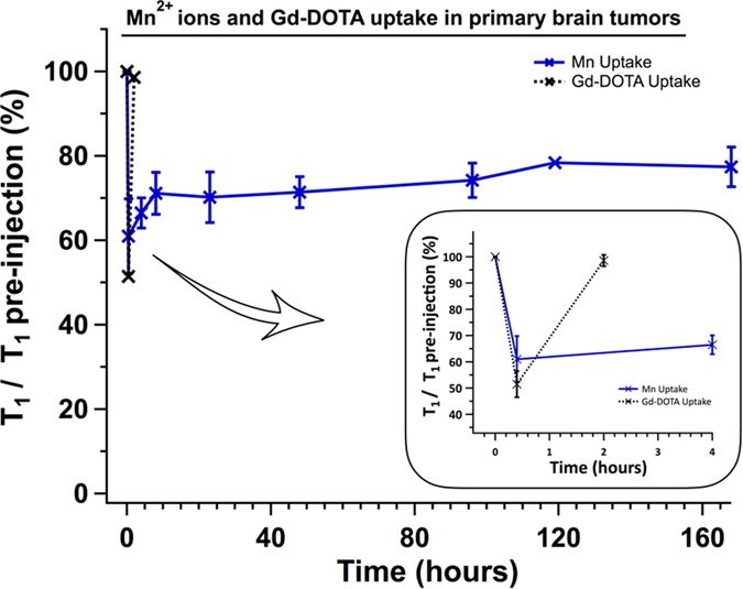
Although MEMRI (Manganese Enhanced MRI) informations were obtained on primary tumors in small animals, MEMRI data on metastases are lacking. Thus, our goal was to determine if 3D Look-Locker T1 mapping was an efficient method to evaluate Mn ions transport in brain metastases in vivo. The high spatial resolution in 3D (156 × 156 × 218 μm) of the sequence enabled to detect metastases of 0.3 mm. In parallel, the T1 quantitation enabled to distinguish three populations of MDA-MB-231 derived brain metastases after MnCl2 intravenous injection: one with a healthy blood-tumor barrier that did not internalize Mn ions, and two others, which T1 shortened drastically by 54.2% or 24%. Subsequent scans of the mice, enabled by the fast acquisition (23 min), demonstrated that these T1 reached back their pre-injection values in 24 h. Contrarily to metastases, the T1 of U87-MG glioma remained 26.2% shorter for one week. In vitro results supported the involvement of the Transient Receptor Potential channels and the Calcium-Sensing Receptor in the uptake and efflux of Mn ions, respectively. This study highlights the ability of the 3D Look-Locker T1 mapping sequence to study heterogeneities (i) amongst brain metastases and (ii) between metastases and glioma regarding Mn transport.
虽然在小动物的原发性肿瘤中获得了 MEMRI(锰增强 MRI)信息,但缺乏转移瘤的 MEMRI 数据。因此,我们的目标是确定 3D Look-Locker T1 映射是否是评估体内脑转移瘤中锰离子转运的有效方法。该序列在 3D 中的高空间分辨率(156×156×218μm)能够检测到 0.3mm 的转移灶。同时,T1 定量能够区分静脉注射 MnCl2 后三种 MDA-MB-231 衍生的脑转移瘤:一种血脑屏障健康,不摄取锰离子,另外两种 T1 分别缩短 54.2%或 24%。通过快速采集(23 分钟)对小鼠进行后续扫描,证明这些 T1 在 24 小时内恢复到注射前的水平。与转移瘤相反,U87-MG 神经胶质瘤的 T1 在一周内仍然缩短了 26.2%。体外结果支持瞬时受体电位通道和钙敏感受体分别参与锰离子的摄取和外排。这项研究强调了 3D Look-Locker T1 映射序列研究脑转移瘤之间(i)和转移瘤与神经胶质瘤之间(ii)锰转运异质性的能力。