Quantitative Light imaging Laboratory, Department of Electrical and Computer Engineering, Beckman Institute for Advanced Science and Technology, University of Illinois at Urbana-Champaign, 405 N. Matthews Avenue, Urbana, IL 61801, USA.
Translational Biophotonics Laboratory, Department of Biomedical Engineering, Ulsan National Institute of Science and Technology, 50 UNIST-gil, Ulsan 44919, Republic of Korea.
Sci Rep. 2016 Dec 23;6:39667. doi: 10.1038/srep39667.
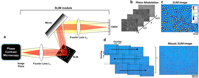
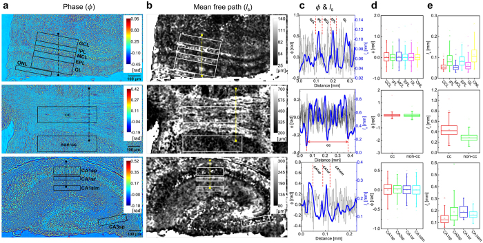
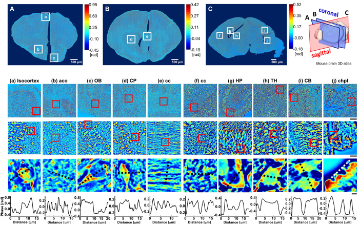
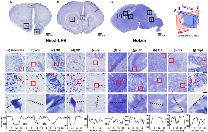
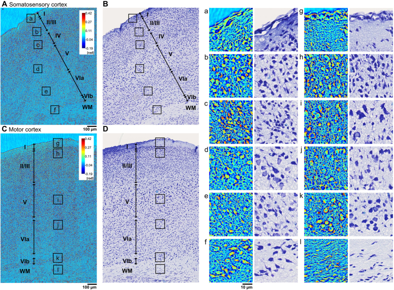
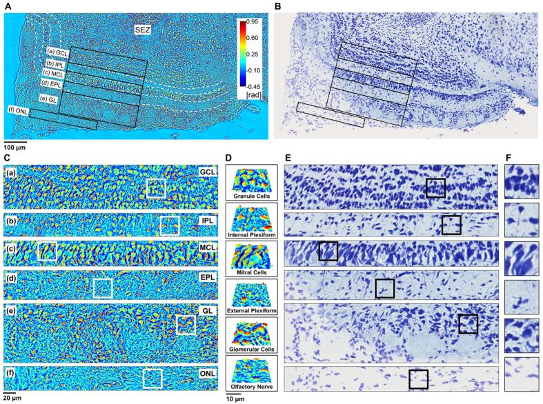
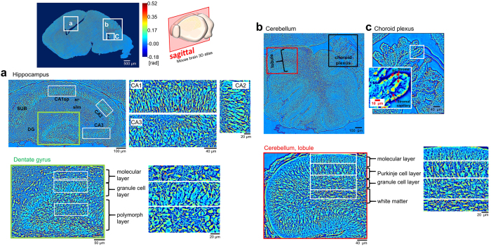
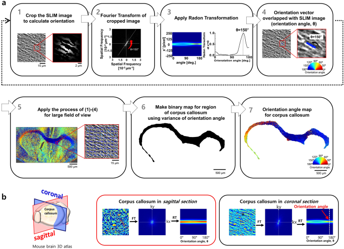
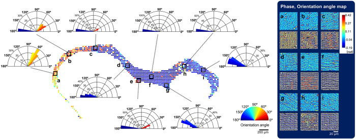
Brain connectivity spans over broad spatial scales, from nanometers to centimeters. In order to understand the brain at multi-scale, the neural network in wide-field has been visualized in detail by taking advantage of light microscopy. However, the process of staining or addition of fluorescent tags is commonly required, and the image contrast is insufficient for delineation of cytoarchitecture. To overcome this barrier, we use spatial light interference microscopy to investigate brain structure with high-resolution, sub-nanometer pathlength sensitivity without the use of exogenous contrast agents. Combining wide-field imaging and a mosaic algorithm developed in-house, we show the detailed architecture of cells and myelin, within coronal olfactory bulb and cortical sections, and from sagittal sections of the hippocampus and cerebellum. Our technique is well suited to identify laminar characteristics of fiber tract orientation within white matter, e.g. the corpus callosum. To further improve the macro-scale contrast of anatomical structures, and to better differentiate axons and dendrites from cell bodies, we mapped the tissue in terms of its scattering property. Based on our results, we anticipate that spatial light interference microscopy can potentially provide multiscale and multicontrast perspectives of gross and microscopic brain anatomy.
脑连接跨越广泛的空间尺度,从纳米到厘米。为了在多尺度上理解大脑,利用显微镜已经详细地可视化了宽场中的神经网络。然而,通常需要染色或添加荧光标记的过程,并且图像对比度不足以描绘细胞结构。为了克服这个障碍,我们使用空间光干涉显微镜以高分辨率、亚纳米路径长度灵敏度来研究大脑结构,而无需使用外源性对比剂。我们结合宽场成像和内部开发的镶嵌算法,展示了冠状嗅球和皮质切片以及海马和小脑矢状切片中细胞和髓鞘的详细结构。我们的技术非常适合识别白质中纤维束方向的层特征,例如胼胝体。为了进一步提高解剖结构的宏观对比度,更好地区分轴突和树突与细胞体,我们根据组织的散射特性对其进行了映射。基于我们的结果,我们预计空间光干涉显微镜可以为大脑的大体和微观解剖结构提供多尺度和多对比度的视角。