Zeng Huawei, Taussig David P, Cheng Wen-Hsing, Johnson LuAnn K, Hakkak Reza
United States Department of Agriculture, Agricultural Research Service, Grand Forks Human Nutrition Research Center, Grand Forks, ND 58203, USA.
Department of Food Science, Nutrition and Health Promotion, Mississippi State University, Starkville, MS 39762, USA.
Nutrients. 2017 Jan 1;9(1):25. doi: 10.3390/nu9010025.
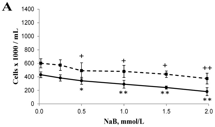
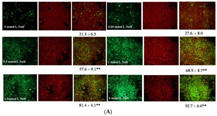
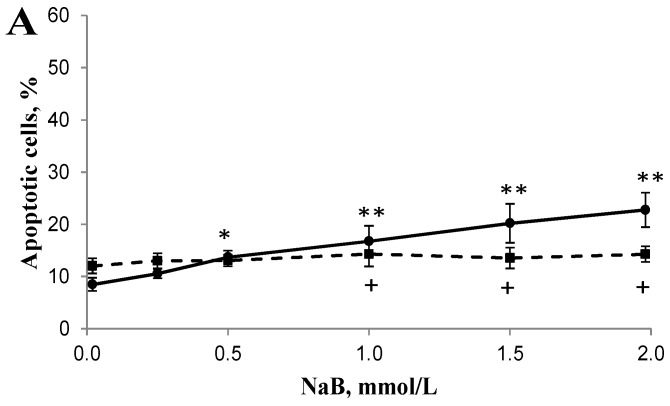
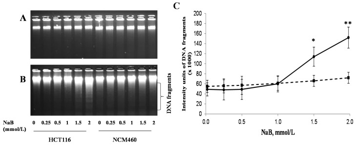
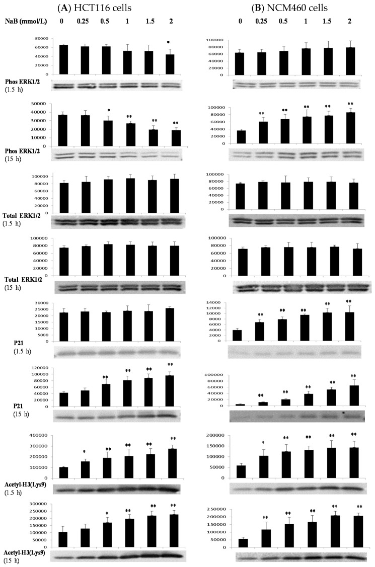
Butyrate, an intestinal microbiota metabolite of dietary fiber, exhibits chemoprevention effects on colon cancer development. However, the mechanistic action of butyrate remains to be determined. We hypothesize that butyrate inhibits cancerous cell proliferation but to a lesser extent in noncancerous cells through regulating apoptosis and cellular-signaling pathways. We tested this hypothesis by exposing cancerous HCT116 or non-cancerous NCM460 colon cells to physiologically relevant doses of butyrate. Cellular responses to butyrate were characterized by Western analysis, fluorescent microscopy, acetylation, and DNA fragmentation analyses. Butyrate inhibited cell proliferation, and led to an induction of apoptosis, genomic DNA fragmentation in HCT116 cells, but to a lesser extent in NCM460 cells. Although butyrate increased H3 histone deacetylation and p21 tumor suppressor expression in both cell types, p21 protein level was greater with intense expression around the nuclei in HCT116 cells when compared with that in NCM460 cells. Furthermore, butyrate treatment increased the phosphorylation of extracellular-regulated kinase 1/2 (p-ERK1/2), a survival signal, in NCM460 cells while it decreased p-ERK1/2 in HCT116 cells. Taken together, the activation of survival signaling in NCM460 cells and apoptotic potential in HCT116 cells may confer the increased sensitivity of cancerous colon cells to butyrate in comparison with noncancerous colon cells.
丁酸是膳食纤维的肠道微生物群代谢产物,对结肠癌的发展具有化学预防作用。然而,丁酸的作用机制仍有待确定。我们假设丁酸通过调节细胞凋亡和细胞信号通路来抑制癌细胞增殖,但对非癌细胞的抑制作用较小。我们通过将癌性HCT116或非癌性NCM460结肠细胞暴露于生理相关剂量的丁酸来验证这一假设。通过蛋白质免疫印迹分析、荧光显微镜检查、乙酰化分析和DNA片段化分析来表征细胞对丁酸的反应。丁酸抑制细胞增殖,并导致HCT116细胞凋亡、基因组DNA片段化,而对NCM460细胞的作用较小。虽然丁酸在两种细胞类型中均增加了H3组蛋白去乙酰化和p21肿瘤抑制因子的表达,但与NCM460细胞相比,HCT116细胞中p21蛋白水平更高,且在细胞核周围有强烈表达。此外,丁酸处理增加了NCM460细胞中细胞外调节激酶1/2(p-ERK1/2)的磷酸化,p-ERK1/2是一种生存信号,而在HCT116细胞中则降低了p-ERK1/2。综上所述,与非癌性结肠细胞相比,NCM460细胞中生存信号的激活和HCT116细胞中的凋亡潜能可能使癌性结肠细胞对丁酸的敏感性增加。