Knight S C, Fryer P, Griffiths S, Harding B, Dixey J, Mansell B
MRC Clinical Research Centre, Harrow, Middlesex, England.
Clin Exp Immunol. 1989 Oct;78(1):19-25.
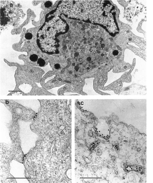
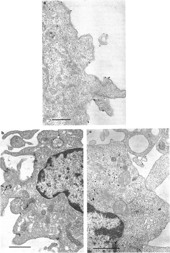
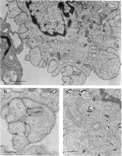
Dendritic cells were enriched from synovial fluids (SF) of patients with inflammatory arthritis and studied by immunogold labelling and electron microscopy for expression of histocompatability antigens of the HLA-D locus. Dendritic cells from SF were larger than most of these from peripheral blood with a more extensive Golgi region and more lysosomes and microfilaments. Class II histocompatability antigens HLA-DR, -DP, -DQ and that labelled by the antibody RFDI were abundant on the dendritic cells. The macrophages in the enriched cells showed labelling for DR but little labelling with the other antibodies. DR, DP and RFDI were often concentrated at areas of contact between dendritic and other cells (other dendritic cells, macrophages or lymphocytes). On incubating labelled cells at 37 degrees C for 30 min many macrophages lost their DR label but dendritic cells always retained some surface label. Some gold labelling DR and DP was found in characteristic channels between the veils and became internalized in membrane-bound structures. A small proportion of the RFDI label internalized in areas resembling coated pits. Less DQ label internalized and appeared on vesicles inside vacuoles. Material bound to different class II molecules may thus be internalized or processed differently by dendritic cells. The presence in inflammatory lesions of large activated dendritic cells with high expression of class II antigens suggests that these cells could be presenting antigen to lymphocytes within the joints.
从炎性关节炎患者的滑液(SF)中富集树突状细胞,并通过免疫金标记和电子显微镜研究HLA - D位点组织相容性抗原的表达。滑液中的树突状细胞比外周血中的大多数树突状细胞更大,具有更广泛的高尔基体区域、更多的溶酶体和微丝。树突状细胞上丰富表达II类组织相容性抗原HLA - DR、- DP、- DQ以及由抗体RFDI标记的抗原。富集细胞中的巨噬细胞显示出DR标记,但与其他抗体的标记很少。DR、DP和RFDI常常集中在树突状细胞与其他细胞(其他树突状细胞、巨噬细胞或淋巴细胞)的接触区域。将标记细胞在37℃孵育30分钟后,许多巨噬细胞失去了它们的DR标记,但树突状细胞总是保留一些表面标记。在面纱之间的特征性通道中发现了一些金标记的DR和DP,并在膜结合结构中内化。一小部分RFDI标记在内化于类似有被小窝的区域。较少的DQ标记内化并出现在液泡内的小泡上。因此,与不同II类分子结合的物质可能被树突状细胞以不同方式内化或处理。具有高表达II类抗原的大型活化树突状细胞在炎性病变中的存在表明,这些细胞可能在向关节内的淋巴细胞呈递抗原。