• 文献检索
  • 文档翻译
  • 深度研究
  • 学术资讯
  • Suppr Zotero 插件Zotero 插件
  • 邀请有礼
  • 套餐&价格
  • 历史记录
应用&插件
Suppr Zotero 插件Zotero 插件浏览器插件Mac 客户端Windows 客户端微信小程序
定价
高级版会员购买积分包购买API积分包
服务
文献检索文档翻译深度研究API 文档MCP 服务
关于我们
关于 Suppr公司介绍联系我们用户协议隐私条款
关注我们

Suppr 超能文献

核心技术专利:CN118964589B侵权必究
粤ICP备2023148730 号-1Suppr @ 2026

文献检索

告别复杂PubMed语法,用中文像聊天一样搜索,搜遍4000万医学文献。AI智能推荐,让科研检索更轻松。

立即免费搜索

文件翻译

保留排版,准确专业,支持PDF/Word/PPT等文件格式,支持 12+语言互译。

免费翻译文档

深度研究

AI帮你快速写综述,25分钟生成高质量综述,智能提取关键信息,辅助科研写作。

立即免费体验

概率性肝脏图谱构建

Probabilistic liver atlas construction.

作者信息

Dura Esther, Domingo Juan, Ayala Guillermo, Marti-Bonmati Luis, Goceri E

机构信息

Department of Informatics, School of Engineering, University of Valencia, Avda. de la Universidad, 46100, Burjasot, Spain.

Department of Statistics and Operations Research, University of Valencia, Avda. Vicent Andrés Estellés, 1, 46100, Burjasot, Spain.

出版信息

Biomed Eng Online. 2017 Jan 13;16(1):15. doi: 10.1186/s12938-016-0305-8.

DOI:10.1186/s12938-016-0305-8
PMID:28086965
原文链接:https://pmc.ncbi.nlm.nih.gov/articles/PMC5237330/
Abstract

BACKGROUND

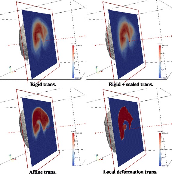
Anatomical atlases are 3D volumes or shapes representing an organ or structure of the human body. They contain either the prototypical shape of the object of interest together with other shapes representing its statistical variations (statistical atlas) or a probability map of belonging to the object (probabilistic atlas). Probabilistic atlases are mostly built with simple estimations only involving the data at each spatial location.

RESULTS

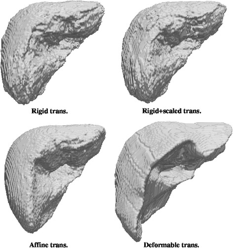
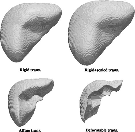
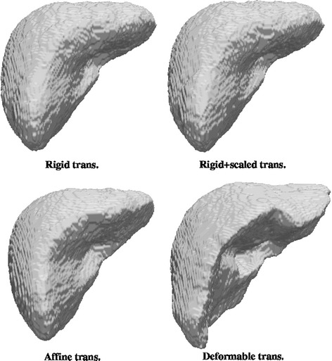
A new method for probabilistic atlas construction that uses a generalized linear model is proposed. This method aims to improve the estimation of the probability to be covered by the liver. Furthermore, all methods to build an atlas involve previous coregistration of the sample of shapes available. The influence of the geometrical transformation adopted for registration in the quality of the final atlas has not been sufficiently investigated. The ability of an atlas to adapt to a new case is one of the most important quality criteria that should be taken into account. The presented experiments show that some methods for atlas construction are severely affected by the previous coregistration step.

CONCLUSION

We show the good performance of the new approach. Furthermore, results suggest that extremely flexible registration methods are not always beneficial, since they can reduce the variability of the atlas and hence its ability to give sensible values of probability when used as an aid in segmentation of new cases.

摘要

背景

解剖图谱是表示人体器官或结构的三维体积或形状。它们包含感兴趣对象的原型形状以及表示其统计变化的其他形状(统计图谱)或属于该对象的概率图(概率图谱)。概率图谱大多仅通过涉及每个空间位置数据的简单估计构建。

结果

提出了一种使用广义线性模型构建概率图谱的新方法。该方法旨在改进肝脏覆盖概率的估计。此外,构建图谱的所有方法都涉及对可用形状样本进行预先配准。用于配准的几何变换对最终图谱质量的影响尚未得到充分研究。图谱适应新病例的能力是应考虑的最重要质量标准之一。所展示的实验表明,一些图谱构建方法受到先前配准步骤的严重影响。

结论

我们展示了新方法的良好性能。此外,结果表明,极其灵活的配准方法并不总是有益的,因为它们可能会降低图谱的可变性,从而降低其在辅助新病例分割时给出合理概率值的能力。

https://cdn.ncbi.nlm.nih.gov/pmc/blobs/bd0f/5237330/7d3d0157e9b9/12938_2016_305_Fig8_HTML.jpg
https://cdn.ncbi.nlm.nih.gov/pmc/blobs/bd0f/5237330/c0d223261a3d/12938_2016_305_Fig1_HTML.jpg
https://cdn.ncbi.nlm.nih.gov/pmc/blobs/bd0f/5237330/f8a7aea95fc2/12938_2016_305_Fig2_HTML.jpg
https://cdn.ncbi.nlm.nih.gov/pmc/blobs/bd0f/5237330/9ded568f5808/12938_2016_305_Fig3_HTML.jpg
https://cdn.ncbi.nlm.nih.gov/pmc/blobs/bd0f/5237330/a8043ef2eaf8/12938_2016_305_Fig4_HTML.jpg
https://cdn.ncbi.nlm.nih.gov/pmc/blobs/bd0f/5237330/0d32a08fd649/12938_2016_305_Fig5_HTML.jpg
https://cdn.ncbi.nlm.nih.gov/pmc/blobs/bd0f/5237330/056261c07688/12938_2016_305_Fig6_HTML.jpg
https://cdn.ncbi.nlm.nih.gov/pmc/blobs/bd0f/5237330/f23fbf2fb5ba/12938_2016_305_Fig7_HTML.jpg
https://cdn.ncbi.nlm.nih.gov/pmc/blobs/bd0f/5237330/7d3d0157e9b9/12938_2016_305_Fig8_HTML.jpg

相似文献

1
Probabilistic liver atlas construction.概率性肝脏图谱构建
Biomed Eng Online. 2017 Jan 13;16(1):15. doi: 10.1186/s12938-016-0305-8.
2
Probabilistic atlas and geometric variability estimation to drive tissue segmentation.用于驱动组织分割的概率图谱与几何变异性估计
Stat Med. 2014 Sep 10;33(20):3576-99. doi: 10.1002/sim.6156. Epub 2014 Apr 2.
3
Medical image segmentation via atlases and fuzzy object models: Improving efficacy through optimum object search and fewer models.通过图谱和模糊对象模型进行医学图像分割:通过优化对象搜索和减少模型提高效率。
Med Phys. 2016 Jan;43(1):401. doi: 10.1118/1.4938577.
4
Automated segmentation of the prostate in 3D MR images using a probabilistic atlas and a spatially constrained deformable model.使用概率图谱和空间约束变形模型对 3D MR 图像中的前列腺进行自动分割。
Med Phys. 2010 Apr;37(4):1579-90. doi: 10.1118/1.3315367.
5
Multi-atlas segmentation of the whole hippocampus and subfields using multiple automatically generated templates.使用多个自动生成的模板对整个海马体及其子区进行多图谱分割。
Neuroimage. 2014 Nov 1;101:494-512. doi: 10.1016/j.neuroimage.2014.04.054. Epub 2014 Apr 29.
6
Segmentation of liver and spleen based on computational anatomy models.基于计算解剖模型的肝脏和脾脏分割
Comput Biol Med. 2015 Dec 1;67:146-60. doi: 10.1016/j.compbiomed.2015.10.007. Epub 2015 Oct 28.
7
Femur statistical atlas construction based on two-level 3D non-rigid registration.基于两级三维非刚性配准的股骨统计图谱构建
Comput Aided Surg. 2009;14(4-6):83-99. doi: 10.3109/10929080903246543.
8
Unbiased diffeomorphic atlas construction for computational anatomy.用于计算解剖学的无偏微分同胚图谱构建
Neuroimage. 2004;23 Suppl 1:S151-60. doi: 10.1016/j.neuroimage.2004.07.068.
9
Unsupervised segmentation, clustering, and groupwise registration of heterogeneous populations of brain MR images.无监督的脑磁共振图像异质人群分割、聚类和组间配准。
IEEE Trans Med Imaging. 2014 Feb;33(2):201-24. doi: 10.1109/TMI.2013.2270114. Epub 2013 Jun 19.
10
Segmentation of 4D cardiac MR images using a probabilistic atlas and the EM algorithm.使用概率图谱和期望最大化算法对4D心脏磁共振图像进行分割。
Med Image Anal. 2004 Sep;8(3):255-65. doi: 10.1016/j.media.2004.06.005.

引用本文的文献

1
Findings from machine learning in clinical medical imaging applications - Lessons for translation to the forensic setting.临床医学影像应用中的机器学习研究结果——对法医学应用的启示。
Forensic Sci Int. 2020 Nov;316:110538. doi: 10.1016/j.forsciint.2020.110538. Epub 2020 Oct 18.
2
Template Creation for High-Resolution Computed Tomography Scans of the Lung in R Software.肺部高分辨率计算机断层扫描模板的 R 软件创建。
Acad Radiol. 2020 Aug;27(8):e204-e215. doi: 10.1016/j.acra.2019.10.030. Epub 2019 Dec 13.

本文引用的文献

1
Statistical shape analysis of the human spleen geometry for probabilistic occupant models.用于概率乘员模型的人体脾脏几何形状的统计形状分析。
J Biomech. 2016 Jun 14;49(9):1540-1546. doi: 10.1016/j.jbiomech.2016.03.027. Epub 2016 Mar 23.
2
Automatic segmentation of the spine by means of a probabilistic atlas with a special focus on ribs suppression. Preliminary results.借助概率图谱对脊柱进行自动分割,特别关注肋骨抑制。初步结果。
Annu Int Conf IEEE Eng Med Biol Soc. 2015;2015:2014-7. doi: 10.1109/EMBC.2015.7318781.
3
Shape-intensity prior level set combining probabilistic atlas and probability map constrains for automatic liver segmentation from abdominal CT images.
基于形状-强度先验水平集的概率图谱和概率图约束的自动肝脏 CT 图像分割方法。
Int J Comput Assist Radiol Surg. 2016 May;11(5):817-26. doi: 10.1007/s11548-015-1332-9. Epub 2015 Dec 8.
4
Automatic localization of the anterior commissure, posterior commissure, and midsagittal plane in MRI scans using regression forests.使用回归森林在MRI扫描中自动定位前连合、后连合和正中矢状面。
IEEE J Biomed Health Inform. 2015 Jul;19(4):1362-74. doi: 10.1109/JBHI.2015.2428672. Epub 2015 Apr 30.
5
How much will linked deformable registrations decrease the quality of multi-atlas segmentation fusions?关联的可变形配准会在多大程度上降低多图谱分割融合的质量?
Radiat Oncol. 2014 Dec 20;9:251. doi: 10.1186/s13014-014-0251-1.
6
Quantitative evaluation of atlas-based high-density diffuse optical tomography for imaging of the human visual cortex.基于图谱的高密度漫射光学层析成像对人类视觉皮层成像的定量评估。
Biomed Opt Express. 2014 Oct 13;5(11):3882-900. doi: 10.1364/BOE.5.003882. eCollection 2014 Nov 1.
7
A statistical geometrical description of the human liver for probabilistic occupant models.用于概率乘员模型的人体肝脏的统计几何描述。
J Biomech. 2014 Nov 28;47(15):3681-8. doi: 10.1016/j.jbiomech.2014.09.031. Epub 2014 Oct 2.
8
Statistical modeling of human liver incorporating the variations in shape, size, and material properties.纳入形状、大小和材料属性变化的人体肝脏统计建模。
Stapp Car Crash J. 2013 Nov;57:285-311. doi: 10.4271/2013-22-0012.
9
Clinical evaluation of multi-atlas based segmentation of lymph node regions in head and neck and prostate cancer patients.基于多图谱的头颈部和前列腺癌患者淋巴结区域分割的临床评估。
Radiat Oncol. 2013 Oct 3;8:229. doi: 10.1186/1748-717X-8-229.
10
Automated abdominal multi-organ segmentation with subject-specific atlas generation.基于特定个体图谱生成的自动腹部多器官分割。
IEEE Trans Med Imaging. 2013 Sep;32(9):1723-30. doi: 10.1109/TMI.2013.2265805. Epub 2013 Jun 3.