Graves Christina L, Li Jian, LaPato Melissa, Shapiro Melanie R, Glover Sarah C, Wallet Mark A, Wallet Shannon M
Department of Oral Biology, College of Dentistry, University of Florida Health Science Center , Gainesville, FL , USA.
Department of Gastroenterology, Hepatology, and Nutrition, College of Medicine, University of Florida Health Science Center , Gainesville, FL , USA.
Front Immunol. 2017 Jan 10;7:679. doi: 10.3389/fimmu.2016.00679. eCollection 2016.
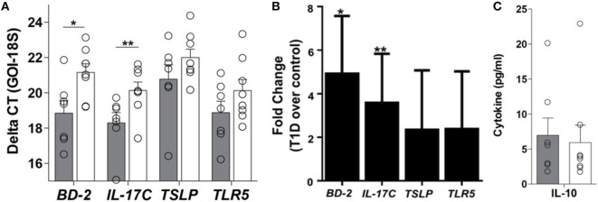
Environmental factors contribute to the initiation, progression, and maintenance of type 1 diabetes (T1D), although a single environmental trigger for disease has not been identified. Studies have documented the contribution of immunity within the gastrointestinal tract (GI) to the expression of autoimmunity at distal sites. Intestinal epithelial cells (IECs) regulate local and systemic immunologic homeostasis through physical and biochemical interactions with innate and adaptive immune populations. We hypothesize that a loss in the tolerance-inducing nature of the GI tract occurs within T1D and is due to altered IECs' innate immune function. As a first step in addressing this hypothesis, we contrasted the global immune microenvironment within the GI tract of individuals with T1D as well as evaluated the IEC-specific effects on adaptive immune cell phenotypes. The soluble and cellular immune microenvironment within the duodenum, the soluble mediator profile of primary IECs derived from the same duodenal tissues, and the effect of the primary IECs' soluble mediator profile on T-cell expansion and polarization were evaluated. Higher levels of IL-17C and beta-defensin 2 (BD-2) mRNA in the T1D-duodenum were observed. Higher frequencies of type 1 innate lymphoid cells (ILC1) and CD8+CXCR3+ T-cells (Tc1) were also observed in T1D-duodenal tissues, concomitant with lower frequencies of type 3 ILC (ILC3) and CD8+CCR6+ T-cells (Tc17). Higher levels of proinflammatory mediators (IL-17C and BD-2) in the absence of similar changes in mediators associated with homeostasis (interleukin 10 and thymic stromal lymphopoietin) were also observed in T1D-derived primary IEC cultures. T1D-derived IEC culture supernatants induced more robust CD8+ T-cell proliferation along with enhanced polarization of Tc1 populations, at the expense of Tc17 polarization, as well as the expansion of CXCR3+CCR6+/- Tregs, indicative of a Th1-like and less regulatory phenotype. These data demonstrate a proinflammatory microenvironment of the T1D-duodenum, whereby IECs have the potential to contribute to the expansion and polarization of innate and adaptive immune cells. Although these data do not discern whether these observations are not simply a consequence of T1D, the data indicate that the T1D-GI tract has the capacity to foster a permissive environment under which autoreactive T-cells could be expanded and polarized.
环境因素对1型糖尿病(T1D)的发病、进展和维持有影响,尽管尚未确定单一的疾病环境触发因素。研究已证明胃肠道(GI)内的免疫对远端部位自身免疫表达的作用。肠上皮细胞(IECs)通过与先天性和适应性免疫细胞群的物理和生化相互作用来调节局部和全身免疫稳态。我们假设,T1D患者胃肠道诱导耐受的特性丧失,这是由于IECs先天性免疫功能改变所致。作为验证这一假设的第一步,我们对比了T1D患者胃肠道内的整体免疫微环境,并评估了IECs对适应性免疫细胞表型的特异性影响。我们评估了十二指肠内的可溶性和细胞免疫微环境、源自相同十二指肠组织的原代IECs的可溶性介质谱,以及原代IECs的可溶性介质谱对T细胞扩增和极化的影响。观察到T1D患者十二指肠中IL-17C和β-防御素2(BD-2)mRNA水平较高。在T1D患者的十二指肠组织中还观察到1型先天性淋巴细胞(ILC1)和CD8+CXCR3+T细胞(Tc1)的频率较高,同时3型ILC(ILC3)和CD8+CCR6+T细胞(Tc17)的频率较低。在源自T1D患者的原代IECs培养物中也观察到促炎介质(IL-17C和BD-2)水平较高,而与稳态相关的介质(白细胞介素10和胸腺基质淋巴细胞生成素)没有类似变化。源自T1D患者的IECs培养上清液诱导更强劲的CD8+T细胞增殖,同时增强Tc1群体的极化,以牺牲Tc17极化以及CXCR3+CCR6+/-调节性T细胞(Tregs)的扩增为代价,这表明呈现出类似Th1的、调节性较弱的表型。这些数据证明了T1D患者十二指肠的促炎微环境,其中IECs有可能促进先天性和适应性免疫细胞的扩增和极化。尽管这些数据无法确定这些观察结果是否不仅仅是T1D的结果,但数据表明T1D患者的胃肠道有能力营造一个宽松的环境,在此环境下自身反应性T细胞可能会扩增和极化。