Terada Makoto, Nakamagoe Kiyotaka, Obara Naoshi, Ogawa Shinichi, Sakamoto Noriaki, Sato Taiki, Nohara Seitaro, Chiba Shigeru, Tamaoka Akira
Department of Neurology, Faculty of Medicine, University of Tsukuba, Japan.
Intern Med. 2017;56(3):363-368. doi: 10.2169/internalmedicine.56.7329. Epub 2017 Feb 1.
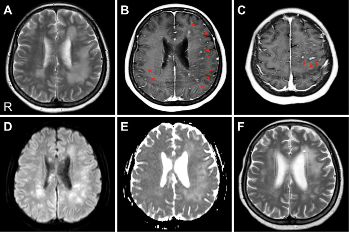
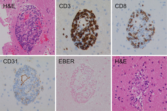
Central nervous system graft-versus-host disease can present quite a diagnostic challenge. We herein present a case of histologically-confirmed chronic graft versus host disease (GVHD) involving the central nervous system that occurred at 19 months after peripheral blood stem cell transplantation. Cranial magnetic resonance imaging showed areas of confluent hyperintensity in the deep/subcortical white matter with multiple punctate and curvilinear gadolinium enhancements, suggesting the disruption of the blood-brain barrier. A brain biopsy revealed perivascular CD3-positive T cell infiltration around the small vessels. We propose that the detection of punctate-enhanced lesions by magnetic resonance imaging may be a useful finding that facilitates the early diagnosis of chronic GVHD involving the central nervous system.
中枢神经系统移植物抗宿主病可能带来相当大的诊断挑战。我们在此报告一例经组织学确诊的慢性移植物抗宿主病(GVHD)累及中枢神经系统的病例,该病例发生在外周血干细胞移植后19个月。头颅磁共振成像显示深部/皮质下白质有融合性高信号区,伴有多个点状和曲线状钆增强,提示血脑屏障破坏。脑活检显示小血管周围有血管周围CD3阳性T细胞浸润。我们认为,磁共振成像检测到点状强化病变可能是一个有助于早期诊断累及中枢神经系统的慢性GVHD的有用发现。