Division of Cardiology, Department of Medicine, UCLA, Los Angeles, CA 90095, USA.
Department of Bioengineering, School of Engineering &Applied Sciences, UCLA, Los Angeles, CA 90095, USA.
Sci Rep. 2017 Feb 6;7:42209. doi: 10.1038/srep42209.
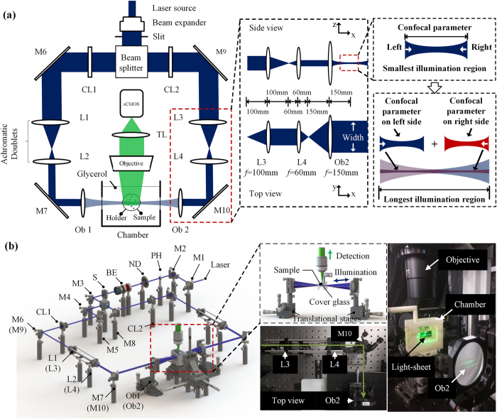
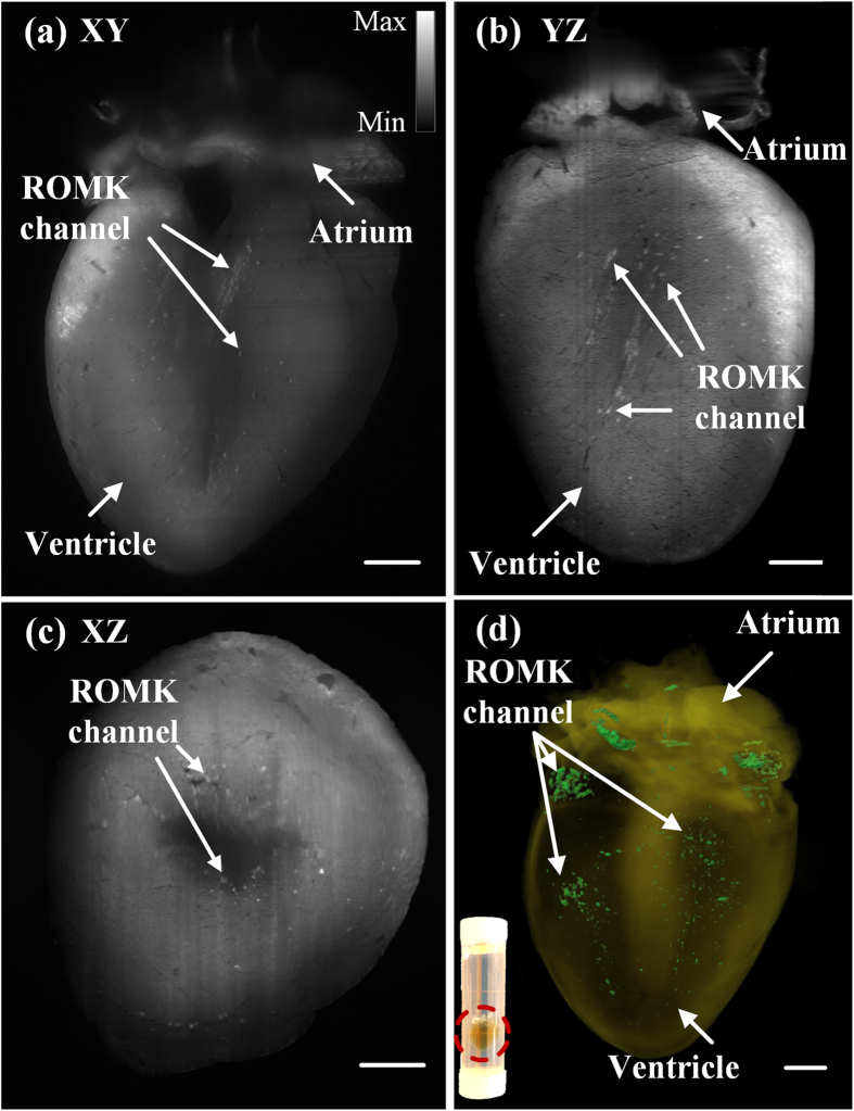
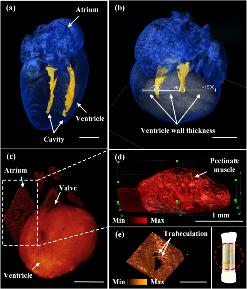
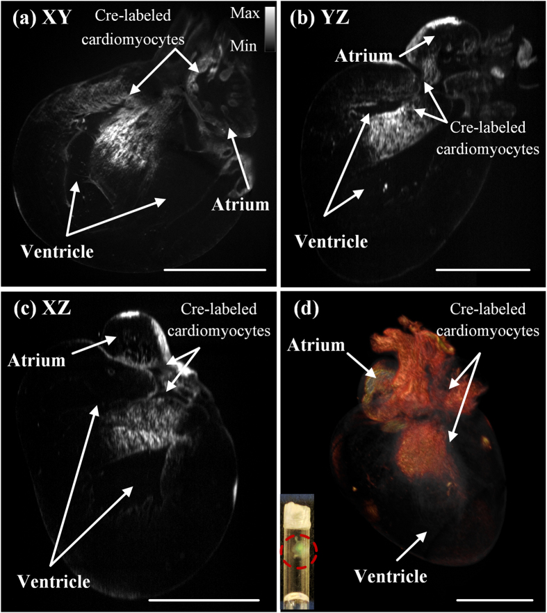
Light-sheet fluorescence microscopy (LSFM) serves to advance developmental research and regenerative medicine. Coupled with the paralleled advances in fluorescence-friendly tissue clearing technique, our cardiac LSFM enables dual-sided illumination to rapidly uncover the architecture of murine hearts over 10 by 10 by 10 mm in volume; thereby allowing for localizing progenitor differentiation to the cardiomyocyte lineage and AAV9-mediated expression of exogenous transmembrane potassium channels with high contrast and resolution. Without the steps of stitching image columns, pivoting the light-sheet and sectioning the heart mechanically, we establish a holistic strategy for 3-dimentional reconstruction of the "digital murine heart" to assess aberrant cardiac structures as well as the spatial distribution of the cardiac lineages in neonates and ion-channels in adults.
光片荧光显微镜 (LSFM) 可用于推进发育研究和再生医学。结合荧光友好型组织透明化技术的平行进展,我们的心脏 LSFM 实现了双侧照明,能够快速揭示体积为 10×10×10mm 的小鼠心脏的结构;从而可以定位祖细胞向心肌细胞谱系的分化,以及 AAV9 介导的外源性跨膜钾通道的表达,具有高对比度和分辨率。我们无需进行图像列拼接、光片旋转和心脏机械切片等步骤,建立了一种用于“数字小鼠心脏”的三维重建的整体策略,以评估新生儿心脏结构的异常以及成年心脏谱系和离子通道的空间分布。