Tallent Jamie, Goodall Stuart, Gibbon Karl C, Hortobágyi Tibor, Howatson Glyn
Department of Sport, Exercise and Rehabilitation, Northumbria UniversityNewcastle-upon-Tyne, UK; School of Sport, Health and Applied Science, St Mary's UniversityTwickenham, UK.
Department of Sport, Exercise and Rehabilitation, Northumbria University Newcastle-upon-Tyne, UK.
Front Physiol. 2017 Feb 7;8:57. doi: 10.3389/fphys.2017.00057. eCollection 2017.
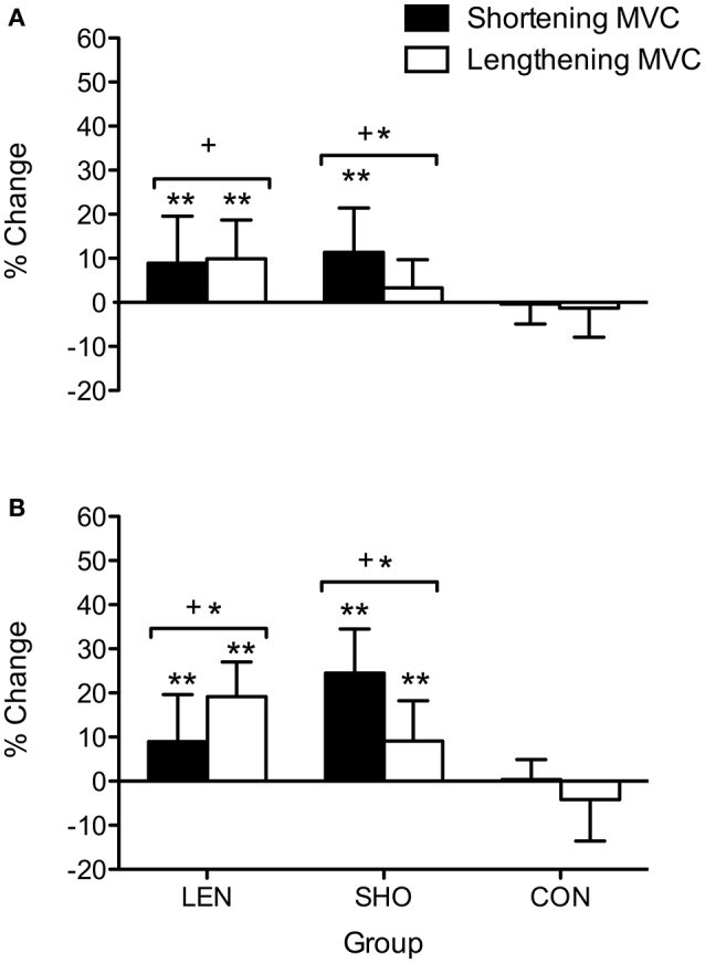
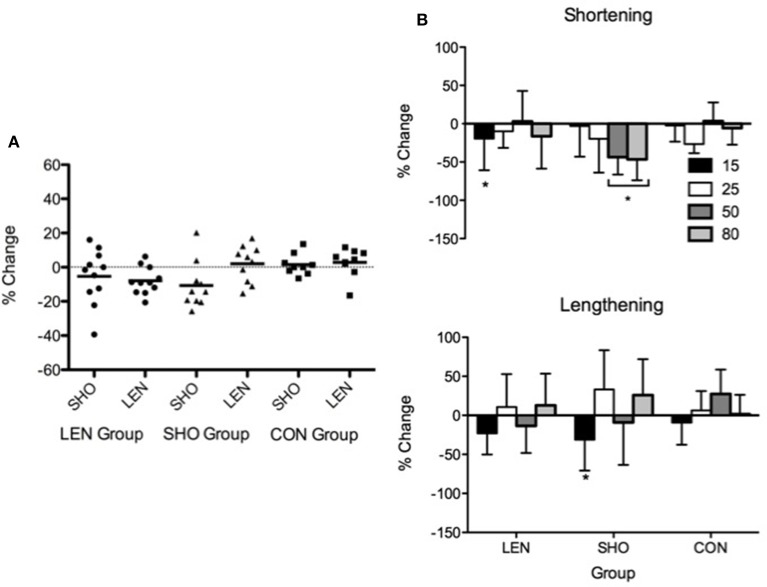
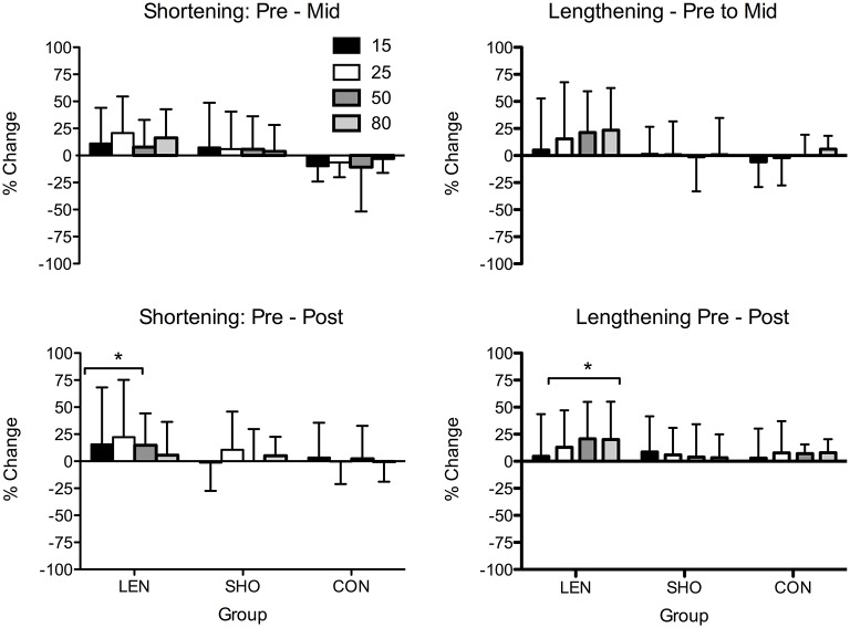
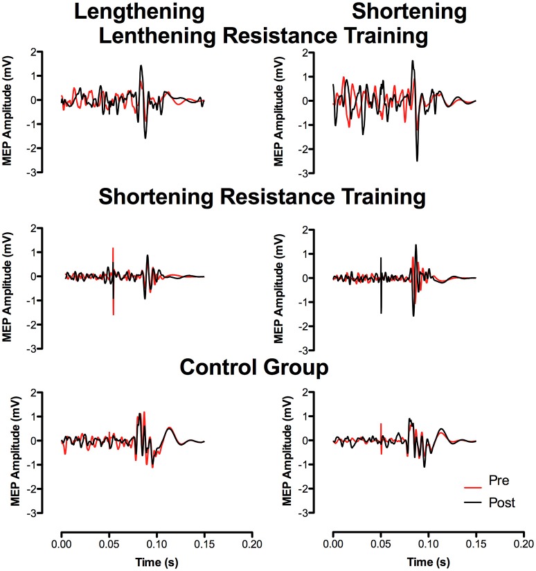
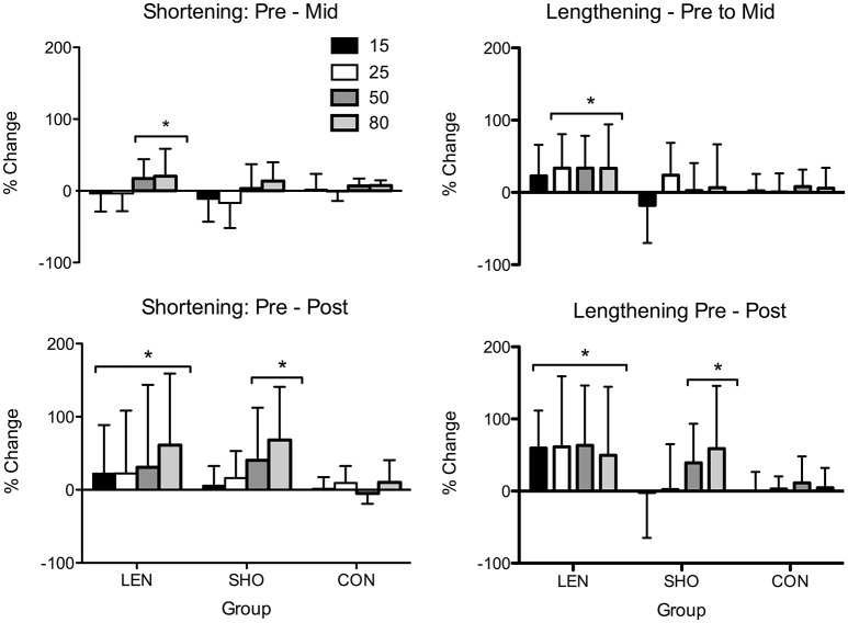
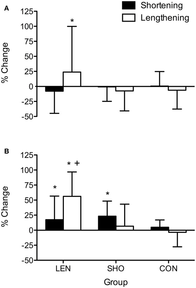
There is a limited understanding of the neurological adaptations responsible for changes in strength following shortening and lengthening resistance training and subsequent detraining. The aim of the study was to investigate differences in corticospinal and spinal responses to resistance training of the tibialis anterior muscle between shortening or lengthening muscle contractions for 4 weeks and after 2 weeks of detraining. Thirty-one untrained individuals were assigned to either shortening or lengthening isokinetic resistance training (4 weeks, 3 days/weeks) or a non-training control group. Transcranial magnetic stimulation and peripheral nerve stimulation (PNS) were used to assess corticospinal and spinal changes, respectively, at pre-, mid-, post-resistance training and post detraining. Greater increases changes ( < 0.01) in MVC were found from the respective muscle contraction training. Motor evoked potentials (expressed relative to background EMG) significantly increased in lengthening resistance training group under contraction intensities ranging from 25 to 80% of the shortening and lengthening contraction intensity ( < 0.01). In the shortening resistance training group increases were only seen at 50 and 80% of both contraction type. Volitional drive (V-wave) showed a greater increase following lengthening resistance training (57%) during maximal lengthening contractions compared to maximal shortening contractions following shortening resistance training (23%; < 0.001). During the detraining period MVC and V-wave did not change ( > 0.05), although MEP amplitude decreased during the detraining period ( < 0.01). No changes in H-reflex were found pre to post resistance training or post detraining. Modulation in V-wave appeared to be contraction specific, whereby greatest increases occurred following lengthening resistance training. Strength and volitional drive is maintained following 2 weeks detraining, however corticospinal excitability appears to decrease when the training stimulus is withdrawn.
目前对于缩短和延长阻力训练以及随后的停训后力量变化所涉及的神经适应性了解有限。本研究的目的是调查在4周的缩短或延长肌肉收缩的胫骨前肌阻力训练以及2周停训后,皮质脊髓和脊髓对阻力训练反应的差异。31名未经训练的个体被分配到缩短或延长等速阻力训练组(4周,每周3天)或非训练对照组。分别在阻力训练前、中、后以及停训后,使用经颅磁刺激和外周神经刺激(PNS)来评估皮质脊髓和脊髓的变化。从各自的肌肉收缩训练中发现最大自主收缩(MVC)有更大的增加变化(<0.01)。在收缩强度为缩短和延长收缩强度的25%至80%范围内,延长阻力训练组的运动诱发电位(相对于背景肌电图表示)显著增加(<0.01)。在缩短阻力训练组中,仅在两种收缩类型的50%和80%时观察到增加。与缩短阻力训练后的最大缩短收缩相比,延长阻力训练后在最大延长收缩期间,意志驱动(V波)显示出更大的增加(57%),而缩短阻力训练后的最大缩短收缩为23%(<0.001)。在停训期间,MVC和V波没有变化(>0.05),尽管在停训期间运动诱发电位幅度下降(<0.01)。在阻力训练前后或停训后未发现H反射有变化。V波的调制似乎具有收缩特异性,延长阻力训练后增加最大。停训2周后力量和意志驱动得以维持,然而当训练刺激撤除时,皮质脊髓兴奋性似乎会降低。