Noda T, Yajima H, Ito Y
Institute for Virus Research, Kyoto University, Japan.
J Virol. 1988 Jan;62(1):313-24. doi: 10.1128/JVI.62.1.313-324.1988.
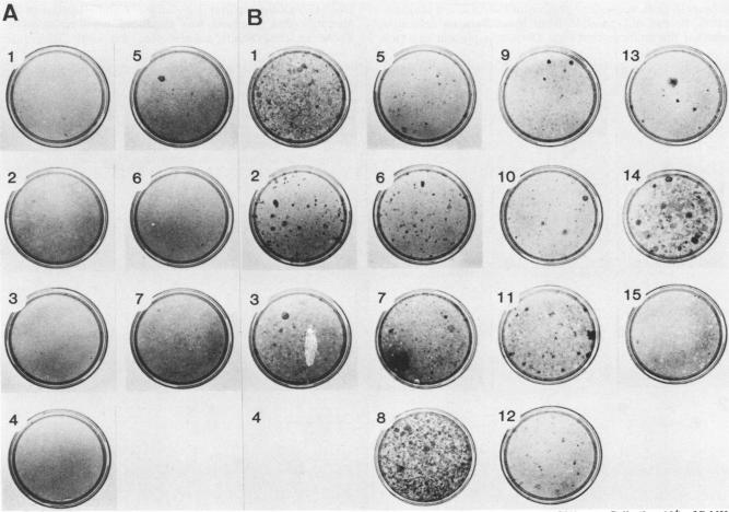
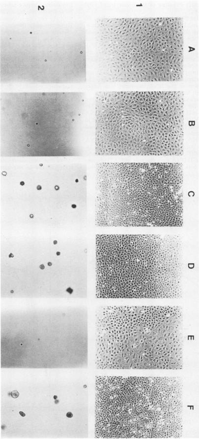
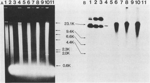
Alteration of the growth properties of the established murine fibroblast cell lines NIH 3T3 and 3Y1 was studied in monolayer cultures and in cells suspended in semisolid medium after introduction of a cloned human papillomavirus type 16 (HPV16) DNA. HPV 16 DNA stimulated both cell lines to grow beyond their saturation densities in monolayer cultures without any apparent morphological changes or tendency to pile up. These cells were also stimulated to grow in soft agar. Since essentially all the cells that received the viral gene were stimulated to grow, the growth-stimulatory activity of HPV16 appeared to be due to the direct effect of a viral gene function. The NIH 3T3 cells showed an additional change in growth properties upon prolonged incubation of dense monolayers of cells containing the HPV16 DNA; morphologically recognizable dense foci appeared at a frequency of about 10(-3). These cells, when cloned from the foci, grew more rapidly in soft agar than the parental cells and were morphologically transformed. In other words, there were two sequential steps in cell transformation induced by HPV16. Practically all the viral DNAs were present in the cells as large rearranged multimers and were integrated into host chromosomal DNA. There was no obvious difference in the state of viral DNA in the cells of the original clone or the three subclones derived from it as dense foci. There was no difference in the amount or the number of viral RNA species expressed in the cells at these two stages. The secondary changes in the growth properties of NIH 3T3 cells appear to be due to some cellular alterations.
在引入克隆的人乳头瘤病毒16型(HPV16)DNA后,研究了已建立的小鼠成纤维细胞系NIH 3T3和3Y1在单层培养物以及悬浮于半固体培养基中的细胞中的生长特性改变。HPV 16 DNA刺激这两种细胞系在单层培养物中生长超过其饱和密度,且无明显形态变化或堆积倾向。这些细胞在软琼脂中也被刺激生长。由于基本上所有接受病毒基因的细胞都被刺激生长,HPV16的生长刺激活性似乎是由于病毒基因功能的直接作用。在含有HPV16 DNA的致密单层细胞长时间孵育后,NIH 3T3细胞的生长特性出现了额外变化;形态上可识别的致密灶以约10^(-3)的频率出现。从这些灶中克隆的细胞在软琼脂中比亲代细胞生长得更快,并且发生了形态转化。换句话说,HPV16诱导的细胞转化有两个连续步骤。实际上所有病毒DNA都以大的重排多聚体形式存在于细胞中,并整合到宿主染色体DNA中。在原始克隆细胞或从致密灶衍生的三个亚克隆细胞中,病毒DNA的状态没有明显差异。在这两个阶段,细胞中表达的病毒RNA种类的数量或种类没有差异。NIH 3T3细胞生长特性的二次变化似乎是由于一些细胞改变。