Fonseca Maria I, Chu Shu-Hui, Hernandez Michael X, Fang Melody J, Modarresi Lila, Selvan Pooja, MacGregor Grant R, Tenner Andrea J
Department of Molecular Biology and Biochemistry, University of California, Irvine, Irvine, CA, 92697, USA.
Department of Pathology and Laboratory Medicine, University of California, Irvine School of Medicine, Irvine, CA, 92697, USA.
J Neuroinflammation. 2017 Mar 6;14(1):48. doi: 10.1186/s12974-017-0814-9.
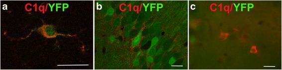
The complement cascade not only provides protection from infection but can also mediate destructive inflammation. Complement is also involved in elimination of neuronal synapses which is essential for proper development, but can be detrimental during aging and disease. C1q, required for several of these complement-mediated activities, is present in the neuropil, microglia, and a subset of interneurons in the brain.
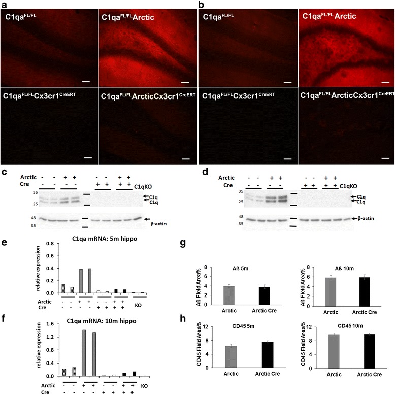
To identify the source(s) of C1q in the brain, the C1qa gene was selectively inactivated in the microglia or Thy-1 neurons in both wild type mice and a mouse model of Alzheimer's disease (AD), and C1q synthesis assessed by immunohistochemistry, QPCR, and western blot analysis.
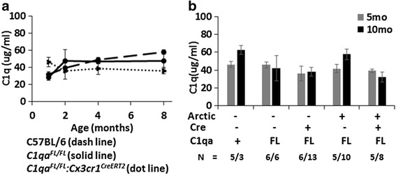
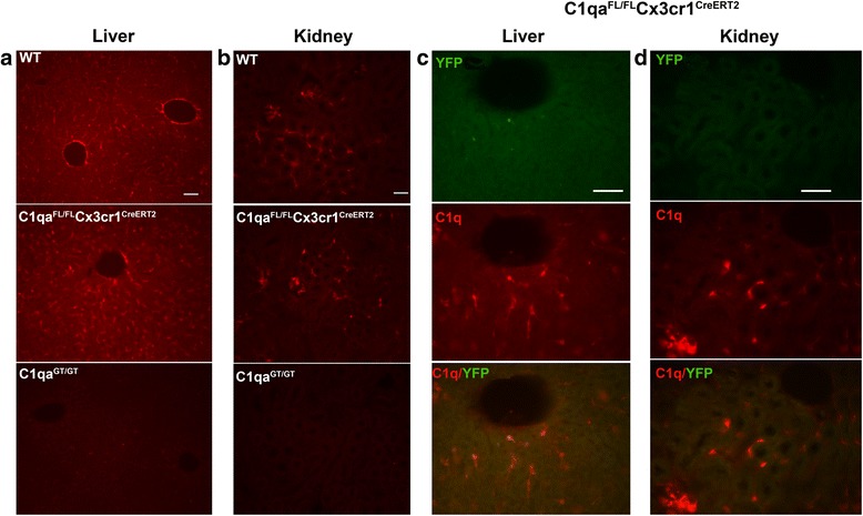
While C1q expression in the brain was unaffected after inactivation of C1qa in Thy-1 neurons, the brains of C1qa :Cx3cr1 mice in which C1qa was ablated in microglia were devoid of C1q with the exception of limited C1q in subsets of interneurons. Surprisingly, this loss of C1q occurred even in the absence of tamoxifen by 1 month of age, demonstrating that Cre activity is tamoxifen-independent in microglia in Cx3cr1 mice. C1q expression in C1qa : Cx3cr1 mice continued to decline and remained almost completely absent through aging and in AD model mice. No difference in C1q was detected in the liver or kidney from C1qa : Cx3cr1 mice relative to controls, and C1qa : Cx3cr1 mice had minimal, if any, reduction in plasma C1q.
Thus, microglia, but not neurons or peripheral sources, are the dominant source of C1q in the brain. While demonstrating that the Cx3cr1 deleter cannot be used for adult-induced deletion of genes in microglia, the model described here enables further investigation of physiological roles of C1q in the brain and identification of therapeutic targets for the selective control of complement-mediated activities contributing to neurodegenerative disorders.
补体级联反应不仅能提供抗感染保护,还能介导破坏性炎症。补体也参与神经元突触的清除,这对正常发育至关重要,但在衰老和疾病过程中可能有害。这些补体介导的活动中有几种所需的C1q存在于脑的神经毡、小胶质细胞和一部分中间神经元中。
为了确定脑中C1q的来源,在野生型小鼠和阿尔茨海默病(AD)小鼠模型的小胶质细胞或Thy-1神经元中选择性地使C1qa基因失活,并通过免疫组织化学、定量聚合酶链反应(QPCR)和蛋白质印迹分析评估C1q的合成。
虽然在Thy-1神经元中使C1qa失活后脑中C1q的表达未受影响,但在小胶质细胞中使C1qa缺失的C1qa:Cx3cr1小鼠的脑,除了中间神经元亚群中有有限的C1q外,没有C1q。令人惊讶的是,即使在没有他莫昔芬的情况下,到1月龄时这种C1q的缺失也会发生,这表明在Cx3cr1小鼠的小胶质细胞中Cre活性不依赖于他莫昔芬。C1qa:Cx3cr1小鼠中的C1q表达持续下降,并且在衰老过程中和AD模型小鼠中几乎完全缺失。相对于对照组,在C1qa:Cx3cr1小鼠的肝脏或肾脏中未检测到C1q的差异,并且C1qa:Cx3cr1小鼠血浆C1q即使有减少也极少。
因此,小胶质细胞而非神经元或外周来源是脑中C1q的主要来源。虽然表明Cx3cr1基因敲除小鼠不能用于成年期诱导的小胶质细胞基因缺失,但此处描述的模型能够进一步研究C1q在脑中的生理作用,并确定用于选择性控制导致神经退行性疾病的补体介导活动的治疗靶点。