Alegro Maryana, Theofilas Panagiotis, Nguy Austin, Castruita Patricia A, Seeley William, Heinsen Helmut, Ushizima Daniela M, Grinberg Lea T
Memory and Aging Center, University of California San Francisco, 675 Nelson Rising Lane, San Francisco, CA 94158, USA.
Medical School of the University of São Paulo, Av. Reboucas 381, São Paulo, SP 05401-000, Brazil.
J Neurosci Methods. 2017 Apr 15;282:20-33. doi: 10.1016/j.jneumeth.2017.03.002. Epub 2017 Mar 4.
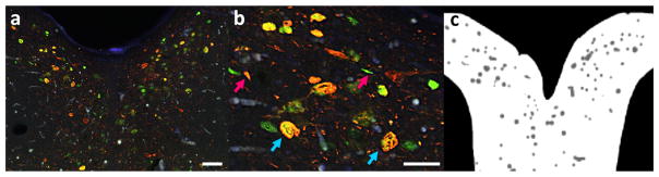
Immunofluorescence (IF) plays a major role in quantifying protein expression in situ and understanding cell function. It is widely applied in assessing disease mechanisms and in drug discovery research. Automation of IF analysis can transform studies using experimental cell models. However, IF analysis of postmortem human tissue relies mostly on manual interaction, often subjected to low-throughput and prone to error, leading to low inter and intra-observer reproducibility. Human postmortem brain samples challenges neuroscientists because of the high level of autofluorescence caused by accumulation of lipofuscin pigment during aging, hindering systematic analyses. We propose a method for automating cell counting and classification in IF microscopy of human postmortem brains. Our algorithm speeds up the quantification task while improving reproducibility.
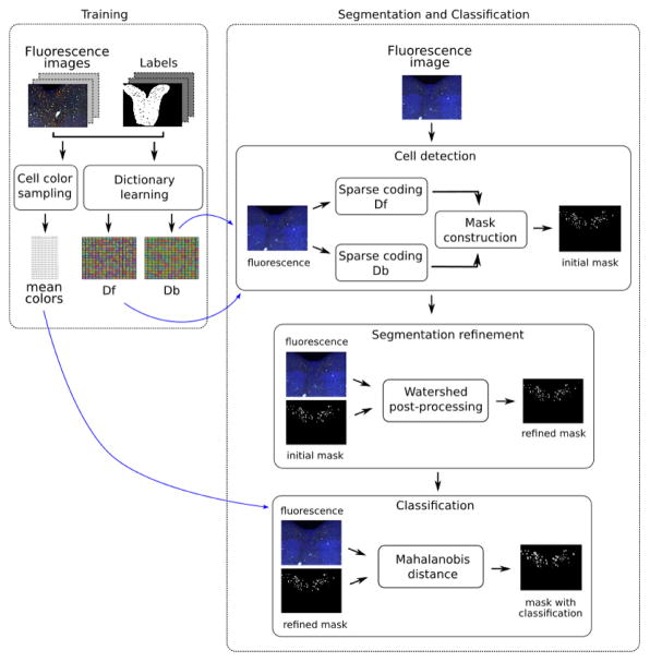
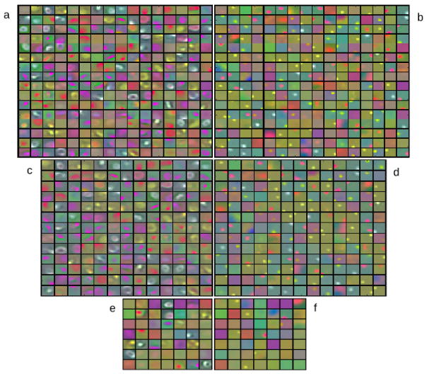
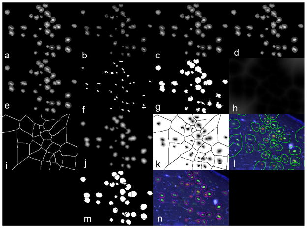
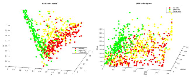
Dictionary learning and sparse coding allow for constructing improved cell representations using IF images. These models are input for detection and segmentation methods. Classification occurs by means of color distances between cells and a learned set.
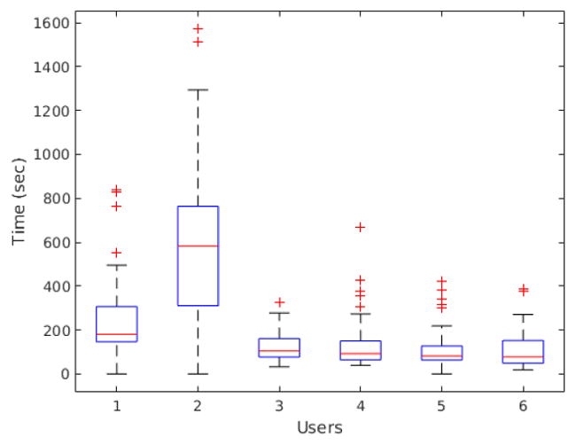
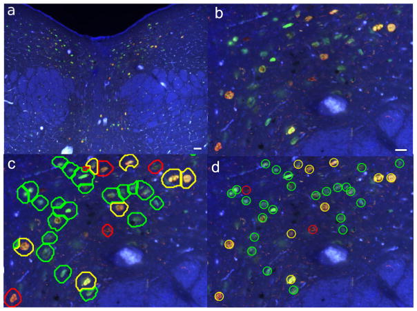
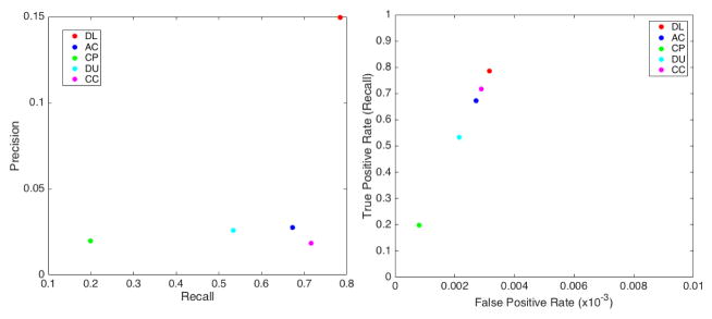
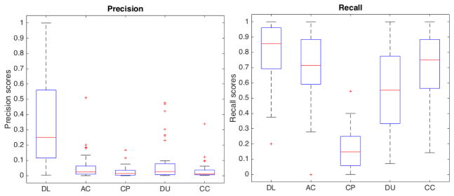
Our method successfully detected and classified cells in 49 human brain images. We evaluated our results regarding true positive, false positive, false negative, precision, recall, false positive rate and F1 score metrics. We also measured user-experience and time saved compared to manual countings.
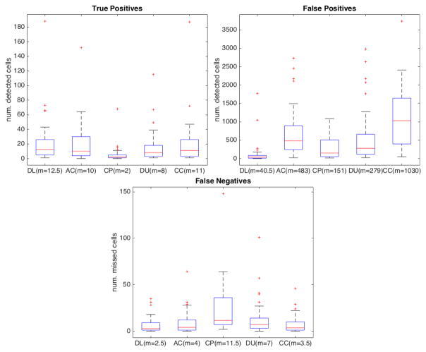
We compared our results to four open-access IF-based cell-counting tools available in the literature. Our method showed improved accuracy for all data samples.
The proposed method satisfactorily detects and classifies cells from human postmortem brain IF images, with potential to be generalized for applications in other counting tasks.
免疫荧光(IF)在原位定量蛋白质表达和理解细胞功能方面发挥着重要作用。它广泛应用于评估疾病机制和药物发现研究。IF分析的自动化可以改变使用实验细胞模型的研究。然而,死后人体组织的IF分析大多依赖人工操作,往往通量低且容易出错,导致观察者间和观察者内的重现性较低。由于衰老过程中脂褐素色素积累导致的高自发荧光水平,人类死后大脑样本给神经科学家带来了挑战,阻碍了系统分析。我们提出了一种用于自动化人类死后大脑IF显微镜下细胞计数和分类的方法。我们的算法在提高重现性的同时加快了定量任务的速度。
字典学习和稀疏编码允许使用IF图像构建改进的细胞表示。这些模型作为检测和分割方法的输入。通过细胞与学习集之间的颜色距离进行分类。
我们的方法成功地在49张人类大脑图像中检测并分类了细胞。我们根据真阳性、假阳性、假阴性、精度、召回率、假阳性率和F1分数指标评估了我们的结果。我们还测量了与手动计数相比的用户体验和节省的时间。
我们将我们的结果与文献中可用的四种基于IF的开放获取细胞计数工具进行了比较。我们的方法对所有数据样本都显示出更高的准确性。
所提出的方法能够令人满意地从人类死后大脑IF图像中检测并分类细胞,有潜力推广到其他计数任务的应用中。