He Wei, Wang Xiao-Yu, Shi Hong, Bai Wan-Zhu, Cheng Bin, Su Yang-Shuai, Yu Xiao-Chun, Jing Xiang-Hong, Zhu Bing
Institute of Acupuncture and Moxibustion, China Academy of Chinese Medical Sciences, Nanxiaojie 16, Dongzhimennei, Beijing, 100700, China.
The Affiliated Hospital, Shandong University of Traditional Chinese Medicine, No. 42, Wenhuaxi Road, Jinan, 250014, China.
BMC Complement Altern Med. 2017 Mar 7;17(1):141. doi: 10.1186/s12906-017-1580-z.
In acupuncture practice, the most important step is to confirm the location of a sensitized acupoint which reflects a diagnosis and can be stimulated with a specialized needle to treat the disease. Abnormal symptoms such as hyperalgesia or allodynia at the sensitized acupoints in patients with visceral disorders are considered to be in relation with referred pain and neurogenic inflammation. Yet, limited study has investigated the cutaneous neurochemical changes of the sensitized acuponits.
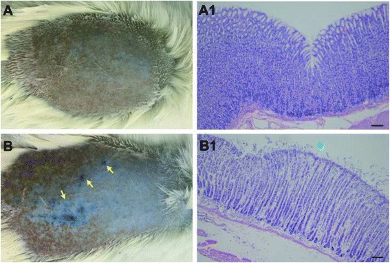
The resent study developed an animal model of gastric mucosal injury (GMI) by HCl administered into the stomach of the rats. Evans Blue (EB) dye was applied by injection of tail vein after mucosal damage to observe the neurogenic plasma extravasation dots in the skin of the rats. The EB dots extravagated in the skin were compared with locations of acupoints. Immnohistochemistry analysis was used to detect the expression of calcitonin gene-related peptide (CGRP)- or substance P (SP)-labeled nerve fibers, histamine (HA)-, serotonin (5-HT)-, and tryptase-labeled cells in EB dots. Images were recorded and analyzed by Confocal imaging system and Olympus Image Processing Software.
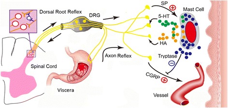
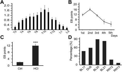
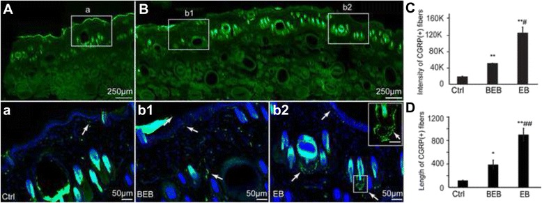
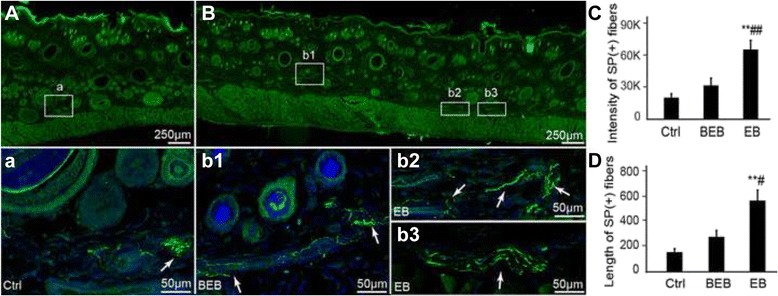
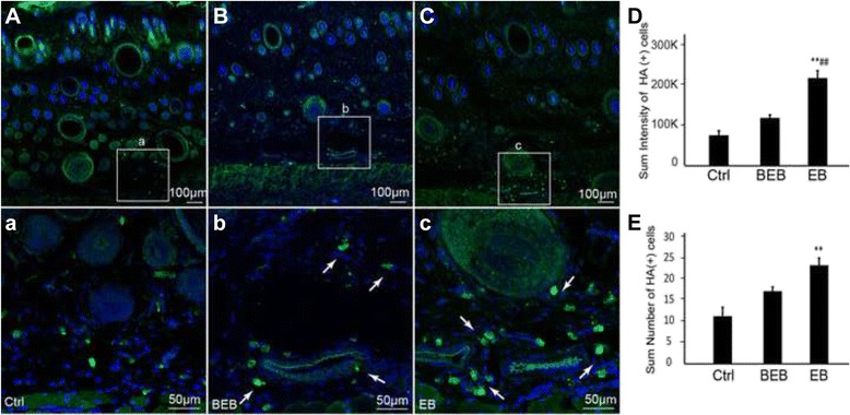
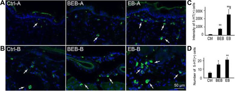
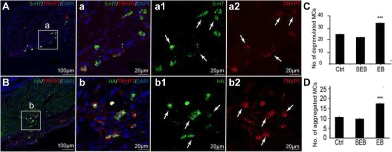
The results showed that GMI resulted in neurogenic plasma extravasation in the skin of the acupoints over the back and abdomen, which mostly occurred in the T9-11 dermatomere. The EB extravasation dots appeared after GMI and disappeared gradually during the natural self-recovery of the gastric mucosa. More SP and CGRP positive nerve fibers were distributed in EB dots than that in regions beside EB dots and in the control, mostly distributed in the nerve fibers around both the vessels and root of hair follicle. Mast cells also aggregated and degranulated to release algogenic substances of 5-HT and HA around the vessels in areas of the EB dots.
Our results indicates that the mechanism of EB extravasation in the skin of the acupoints induced by GMI are closely related to neurogenic inflammation, and that the high expression of local allergic substances and nociceptive neuropeptides in the local skin including SP, CGRP, HA, 5-HT, and mast cell tryptase may be the underlying mechanism of the acupoint sensitization.
在针灸实践中,最重要的步骤是确定敏化穴位的位置,该穴位反映诊断结果,并可用特制针刺激以治疗疾病。内脏疾病患者敏化穴位处的异常症状,如痛觉过敏或异常性疼痛,被认为与牵涉痛和神经源性炎症有关。然而,对敏化穴位皮肤神经化学变化的研究有限。
本研究通过向大鼠胃内注射盐酸建立胃黏膜损伤(GMI)动物模型。黏膜损伤后经尾静脉注射伊文思蓝(EB)染料,观察大鼠皮肤中的神经源性血浆外渗点。将皮肤中出现的EB点与穴位位置进行比较。采用免疫组织化学分析检测EB点中降钙素基因相关肽(CGRP)或P物质(SP)标记的神经纤维、组胺(HA)、5-羟色胺(5-HT)和类胰蛋白酶标记细胞的表达。通过共聚焦成像系统和奥林巴斯图像处理软件记录并分析图像。
结果显示,GMI导致背部和腹部穴位皮肤出现神经源性血浆外渗,主要发生在T9 - 11皮节。GMI后出现EB外渗点,在胃黏膜自然自我恢复过程中逐渐消失。与EB点周围区域及对照组相比,EB点中有更多的SP和CGRP阳性神经纤维分布,大多分布在血管和毛囊根部周围的神经纤维中。肥大细胞也在EB点区域的血管周围聚集并脱颗粒,释放5-HT和HA等致痛物质。
我们的结果表明,GMI诱导的穴位皮肤EB外渗机制与神经源性炎症密切相关,局部皮肤中SP、CGRP、HA、5-HT和肥大细胞类胰蛋白酶等局部过敏物质和伤害性神经肽的高表达可能是穴位敏化的潜在机制。