Martin Neil R W, Aguilar-Agon Kathyrn, Robinson George P, Player Darren J, Turner Mark C, Myers Stephen D, Lewis Mark P
School of Sport, Exercise and Health Sciences, Loughborough University, Loughborough, UK.
Department of Sport and Exercise Sciences, University of Chichester, Chichester, UK.
J Cell Biochem. 2017 Sep;118(9):2599-2605. doi: 10.1002/jcb.25982. Epub 2017 May 15.
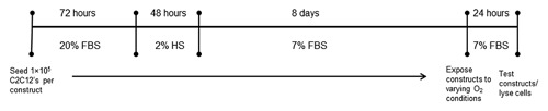
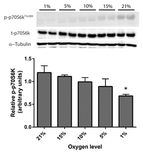
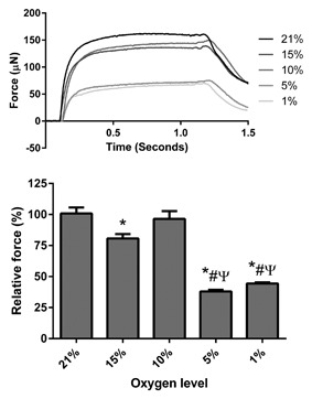
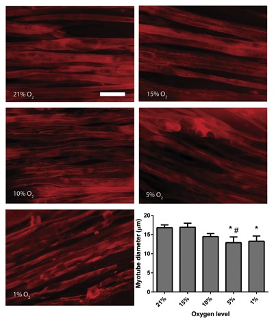
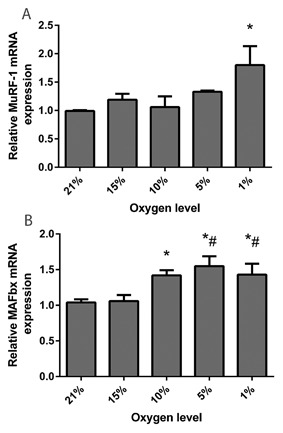
Contemporary tissue engineered skeletal muscle models display a high degree of physiological accuracy compared with native tissue, and therefore may be excellent platforms to understand how various pathologies affect skeletal muscle. Chronic obstructive pulmonary disease (COPD) is a lung disease which causes tissue hypoxia and is characterized by muscle fiber atrophy and impaired muscle function. In the present study we exposed engineered skeletal muscle to varying levels of oxygen (O ; 21-1%) for 24 h in order to see if a COPD like muscle phenotype could be recreated in vitro, and if so, at what degree of hypoxia this occurred. Maximal contractile force was attenuated in hypoxia compared to 21% O ; with culture at 5% and 1% O causing the most pronounced effects with 62% and 56% decrements in force, respectively. Furthermore at these levels of O , myotubes within the engineered muscles displayed significant atrophy which was not seen at higher O levels. At the molecular level we observed increases in mRNA expression of MuRF-1 only at 1% O whereas MAFbx expression was elevated at 10%, 5%, and 1% O . In addition, p70S6 kinase phosphorylation (a downstream effector of mTORC1) was reduced when engineered muscle was cultured at 1% O , with no significant changes seen above this O level. Overall, these data suggest that engineered muscle exposed to O levels of ≤5% adapts in a manner similar to that seen in COPD patients, and thus may provide a novel model for further understanding muscle wasting associated with tissue hypoxia. J. Cell. Biochem. 118: 2599-2605, 2017. © 2017 The Authors. Journal of Cellular Biochemistry Published by Wiley Periodicals, Inc.
与天然组织相比,当代组织工程化骨骼肌模型具有高度的生理准确性,因此可能是了解各种病理状况如何影响骨骼肌的理想平台。慢性阻塞性肺疾病(COPD)是一种导致组织缺氧的肺部疾病,其特征为肌纤维萎缩和肌肉功能受损。在本研究中,我们将工程化骨骼肌暴露于不同水平的氧气(O₂;21%-1%)中24小时,以观察是否能在体外重现类似COPD的肌肉表型,若能重现,是在何种缺氧程度下发生的。与21% O₂相比,缺氧条件下最大收缩力减弱;在5%和1% O₂培养时效果最为显著,力分别下降了62%和56%。此外,在这些O₂水平下,工程化肌肉中的肌管出现了明显萎缩,而在较高O₂水平下未观察到这种情况。在分子水平上,我们仅在1% O₂时观察到MuRF-1的mRNA表达增加,而MAFbx表达在10%、5%和1% O₂时升高。此外,当工程化肌肉在1% O₂培养时,p70S6激酶磷酸化(mTORC1的下游效应器)降低,在该O₂水平以上未观察到显著变化。总体而言,这些数据表明,暴露于≤5% O₂水平的工程化肌肉以类似于COPD患者的方式发生适应性变化,因此可能为进一步了解与组织缺氧相关的肌肉萎缩提供一个新模型。《细胞生物化学杂志》118: 2599 - 2605, 2017。© 2017作者。《细胞生物化学杂志》由威利期刊公司出版。