Sparacia Gianvincenzo, Anastasi Andrea, Speciale Claudia, Agnello Francesco, Banco Aurelia
Gianvincenzo Sparacia, Andrea Anastasi, Claudia Speciale, Francesco Agnello, Aurelia Banco, Department of Radiology, University of Palermo, 90127 Palermo, Italy.
World J Radiol. 2017 Feb 28;9(2):72-78. doi: 10.4329/wjr.v9.i2.72.
To present the typical and atypical magnetic resonance (MR) imaging findings of alcoholic and non-alcoholic Wernicke's encephalopathy.
This study included 7 patients with Wernicke's encephalopathy (2 men, 5 women; mean age, 52.3 years) that underwent brain MR examination between January 2012 and March 2016 in a single institution. Three patients were alcoholics and 4 patients were non-alcoholics. MR protocol included a T2-weighted sequence, a fluid attenuation inversion recovery (FLAIR) sequence, a diffusion-weighted sequence (b = 0 and 1000 s/mm), and a contrast-enhanced MR sequence. All MR images were retrospectively reviewed at baseline and follow-up by two radiologists.
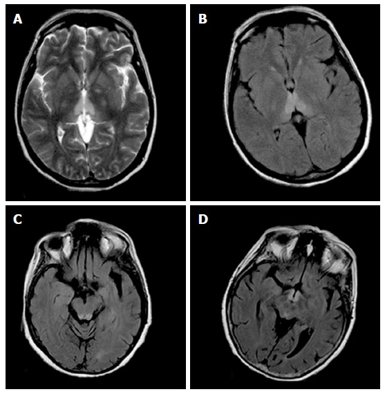
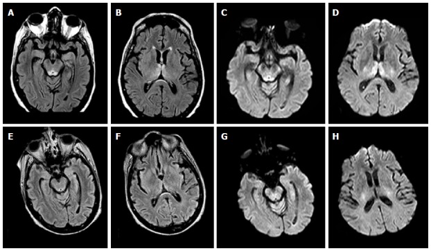
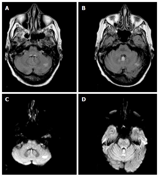
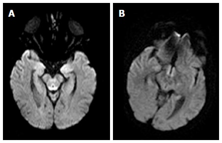
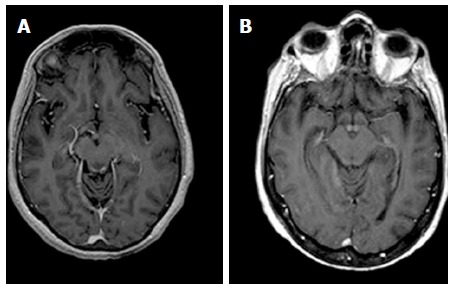
All patients with Wernicke's encephalopathy had bilateral areas showing high signal intensity on both T2-weighted and FLAIR MR images in the typical sites (i.e., the periaqueductal region and the tectal plate). Signal intensity abnormalities in the atypical sites (i.e., the cerebellum and the cerebellar vermis) were seen in 4 patients, all of which had no history of alcohol abuse. Six patients had areas with restricted diffusion in the typical and atypical sites. Four patients had areas showing contrast-enhancement in the typical and atypical sites. Follow-up MR imaging within 6 mo after therapy (intravenous administration of thiamine) was performed in 4 patients, and demonstrated a complete resolution of all the signal intensities abnormalities previously seen in all patients.
MR imaging is valuable in the diagnosis of Wernicke's encephalopathy particularly in patients presenting with atypical clinical symptoms, or with no history of alcohol abuse.
呈现酒精性和非酒精性韦尼克脑病的典型及非典型磁共振(MR)成像表现。
本研究纳入了2012年1月至2016年3月在单一机构接受脑部MR检查的7例韦尼克脑病患者(2例男性,5例女性;平均年龄52.3岁)。其中3例为酗酒者,4例为非酗酒者。MR检查方案包括T2加权序列、液体衰减反转恢复(FLAIR)序列、扩散加权序列(b = 0和1000 s/mm²)以及对比增强MR序列。所有MR图像均由两名放射科医生在基线和随访时进行回顾性分析。
所有韦尼克脑病患者在典型部位(即导水管周围区域和顶盖)的T2加权和FLAIR MR图像上均有双侧高信号区。4例患者在非典型部位(即小脑和小脑蚓部)出现信号强度异常,这些患者均无酗酒史。6例患者在典型和非典型部位有扩散受限区域。4例患者在典型和非典型部位有对比增强区域。4例患者在治疗(静脉注射硫胺素)后6个月内进行了随访MR成像,结果显示所有患者先前出现的信号强度异常均完全消失。
MR成像对韦尼克脑病的诊断具有重要价值,尤其对于临床表现不典型或无酗酒史的患者。