Wiesmann Veit, Bergler Matthias, Palmisano Ralf, Prinzen Martin, Franz Daniela, Wittenberg Thomas
Fraunhofer Institute for Integrated Circuits IIS, Am Wolfsmantel 33, Erlangen, 91058, Germany.
Optical Imaging Centre Erlangen (OICE), Hartmannstraße 14, Erlangen, 91052, Germany.
BMC Bioinformatics. 2017 Mar 18;18(1):176. doi: 10.1186/s12859-017-1591-2.
Manual assessment and evaluation of fluorescent micrograph cell experiments is time-consuming and tedious. Automated segmentation pipelines can ensure efficient and reproducible evaluation and analysis with constant high quality for all images of an experiment. Such cell segmentation approaches are usually validated and rated in comparison to manually annotated micrographs. Nevertheless, manual annotations are prone to errors and display inter- and intra-observer variability which influence the validation results of automated cell segmentation pipelines.
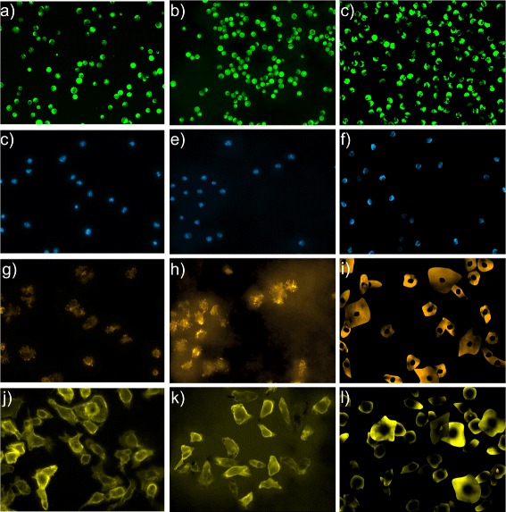
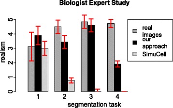
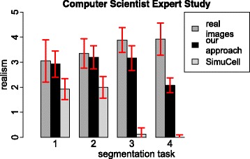
We present a new approach to simulate fluorescent cell micrographs that provides an objective ground truth for the validation of cell segmentation methods. The cell simulation was evaluated twofold: (1) An expert observer study shows that the proposed approach generates realistic fluorescent cell micrograph simulations. (2) An automated segmentation pipeline on the simulated fluorescent cell micrographs reproduces segmentation performances of that pipeline on real fluorescent cell micrographs.
The proposed simulation approach produces realistic fluorescent cell micrographs with corresponding ground truth. The simulated data is suited to evaluate image segmentation pipelines more efficiently and reproducibly than it is possible on manually annotated real micrographs.
对荧光显微图像细胞实验进行人工评估和分析既耗时又繁琐。自动化分割流程能够确保对实验中的所有图像进行高效、可重复的评估和分析,并始终保持高质量。此类细胞分割方法通常通过与人工标注的显微图像进行比较来进行验证和评级。然而,人工标注容易出错,并且存在观察者间和观察者内的差异,这会影响自动化细胞分割流程的验证结果。
我们提出了一种模拟荧光细胞显微图像的新方法,该方法为细胞分割方法的验证提供了客观的真实数据。对细胞模拟进行了两方面的评估:(1)一项专家观察者研究表明,所提出的方法能够生成逼真的荧光细胞显微图像模拟。(2)在模拟荧光细胞显微图像上运行的自动化分割流程能够重现该流程在真实荧光细胞显微图像上的分割性能。
所提出的模拟方法能够生成具有相应真实数据的逼真荧光细胞显微图像。与使用人工标注的真实显微图像相比,模拟数据更适合于对图像分割流程进行高效且可重复的评估。