Kutch Jason J, Labus Jennifer S, Harris Richard E, Martucci Katherine T, Farmer Melissa A, Fenske Sonja, Fling Connor, Ichesco Eric, Peltier Scott, Petre Bogdan, Guo Wensheng, Hou Xiaoling, Stephens Alisa J, Mullins Chris, Clauw Daniel J, Mackey Sean C, Apkarian A Vania, Landis J Richard, Mayer Emeran A
aDivision of Biokinesiology and Physical Therapy, University of Southern California, Los Angeles, CA, USA bG Oppenheimer Center for Neurobiology of Stress and Resilience, Pain and Interoception Network (PAIN), David Geffen School of Medicine at UCLA, Los Angeles, CA, USA cDepartment of Anesthesiology, Chronic Pain and Fatigue Research Center, University of Michigan, Ann Arbor, MI, USA dDepartment of Anesthesiology, Perioperative and Pain Medicine, Division of Pain Medicine, Stanford University Medical Center, Stanford, CA, USA eDepartment of Physiology, Northwestern University, Feinberg School of Medicine, Chicago, IL, USA fNeuroscience Graduate Program, University of Southern California, Los Angeles, CA, USA gFunctional MRI Laboratory, University of Michigan, Ann Arbor, MI, USA hDepartment of Biostatistics and Epidemiology, Perelman School of Medicine at the University of Pennsylvania, Philadelphia, PA, USA iNational Institute of Diabetes and Digestive and Kidney Diseases, NIH, Bethesda, MD, USA.
Pain. 2017 Jun;158(6):1069-1082. doi: 10.1097/j.pain.0000000000000886.
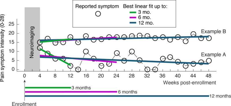
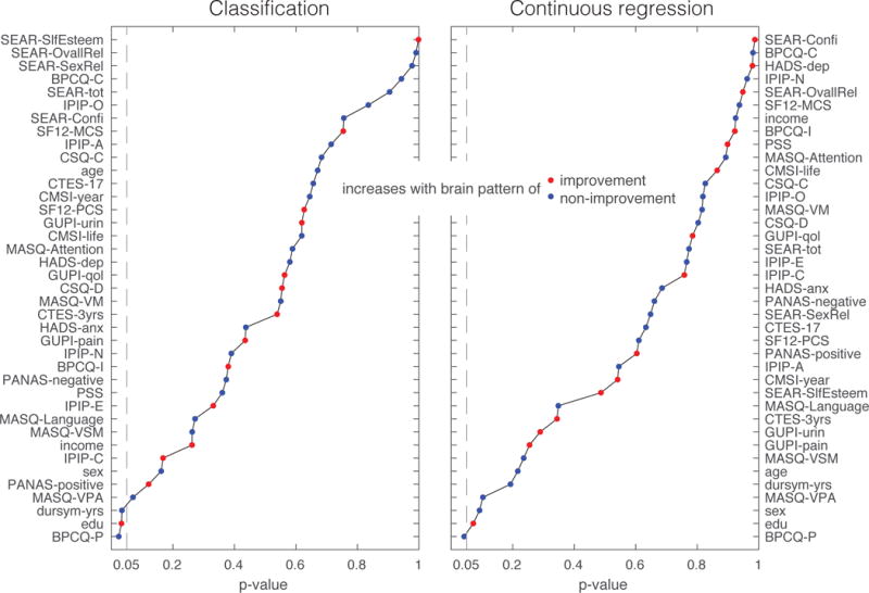
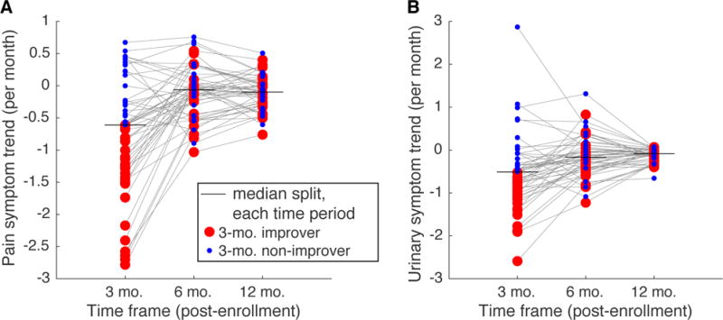
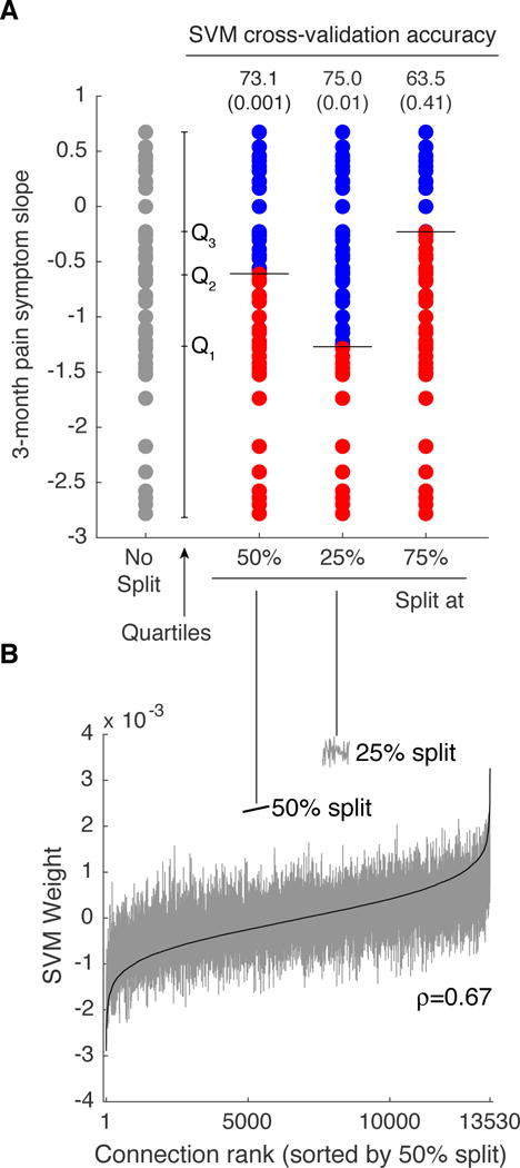
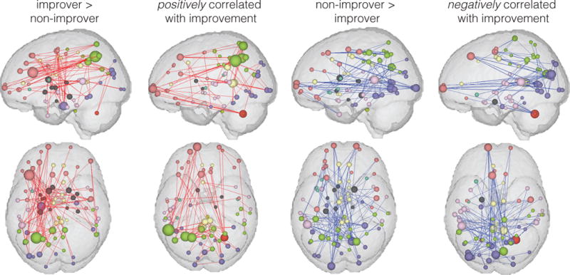
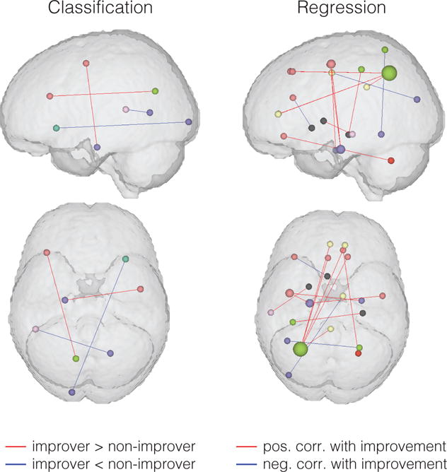
Chronic pain symptoms often change over time, even in individuals who have had symptoms for years. Studying biological factors that predict trends in symptom change in chronic pain may uncover novel pathophysiological mechanisms and potential therapeutic targets. In this study, we investigated whether brain functional connectivity measures obtained from resting-state functional magnetic resonance imaging at baseline can predict longitudinal symptom change (3, 6, and 12 months after scan) in urologic chronic pelvic pain syndrome. We studied 52 individuals with urologic chronic pelvic pain syndrome (34 women, 18 men) who had baseline neuroimaging followed by symptom tracking every 2 weeks for 1 year as part of the Multidisciplinary Approach to the Study of Chronic Pelvic Pain (MAPP) Research Network study. We found that brain functional connectivity can make a significant prediction of short-term (3 month) pain reduction with 73.1% accuracy (69.2% sensitivity and 75.0% precision). In addition, we found that the brain regions with greatest contribution to the classification were preferentially aligned with the left frontoparietal network. Resting-state functional magnetic resonance imaging measures seemed to be less informative about 6- or 12-month symptom change. Our study provides the first evidence that future trends in symptom change in patients in a state of chronic pain may be linked to functional connectivity within specific brain networks.
慢性疼痛症状常常随时间变化,即使是那些已经出现症状多年的个体。研究预测慢性疼痛症状变化趋势的生物学因素,可能会揭示新的病理生理机制和潜在的治疗靶点。在本研究中,我们调查了在基线时通过静息态功能磁共振成像获得的脑功能连接测量值,是否能够预测泌尿外科慢性盆腔疼痛综合征患者的纵向症状变化(扫描后3、6和12个月)。我们研究了52例泌尿外科慢性盆腔疼痛综合征患者(34名女性,18名男性),他们作为慢性盆腔疼痛研究多学科方法(MAPP)研究网络研究的一部分,在基线时进行了神经影像学检查,随后每2周进行一次症状追踪,持续1年。我们发现脑功能连接能够以73.1%的准确率对短期(3个月)疼痛减轻做出显著预测(敏感性为69.2%,精确性为75.0%)。此外,我们发现对分类贡献最大的脑区优先与左侧额顶网络对齐。静息态功能磁共振成像测量值对于6个月或12个月的症状变化似乎信息量较少。我们的研究提供了首个证据,表明处于慢性疼痛状态的患者症状变化的未来趋势可能与特定脑网络内的功能连接有关。