Wang Haibo, Tao Lide, Jin Feng, Gu Hao, Dai Xiaojun, Ni Tengyang, Feng Jun, Ding Yanbing, Xiao Weiming, Qian Yayun, Liu Yanqing
The Affiliated Hospital of Yangzhou University, Yangzhou University, Yangzhou 225000, China.
Clinical Medicine College of Yangzhou University, Yangzhou 225000, China.
Oncotarget. 2017 Jun 13;8(24):39131-39142. doi: 10.18632/oncotarget.16608.
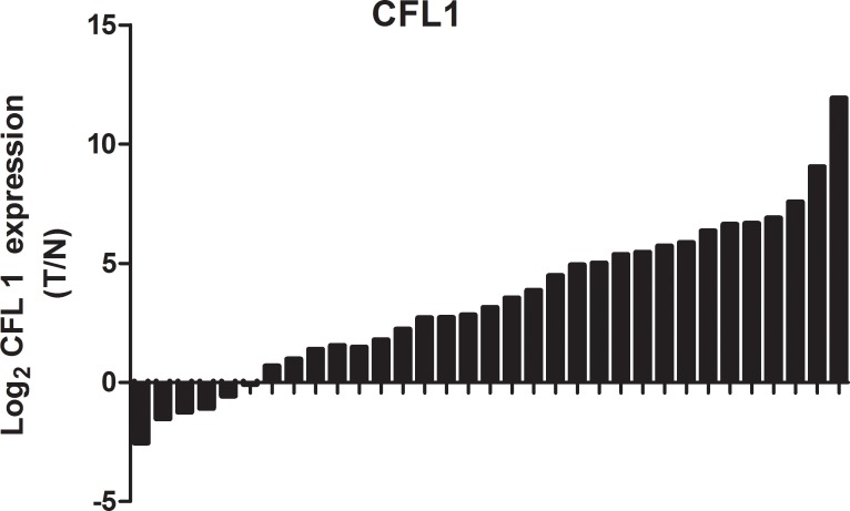
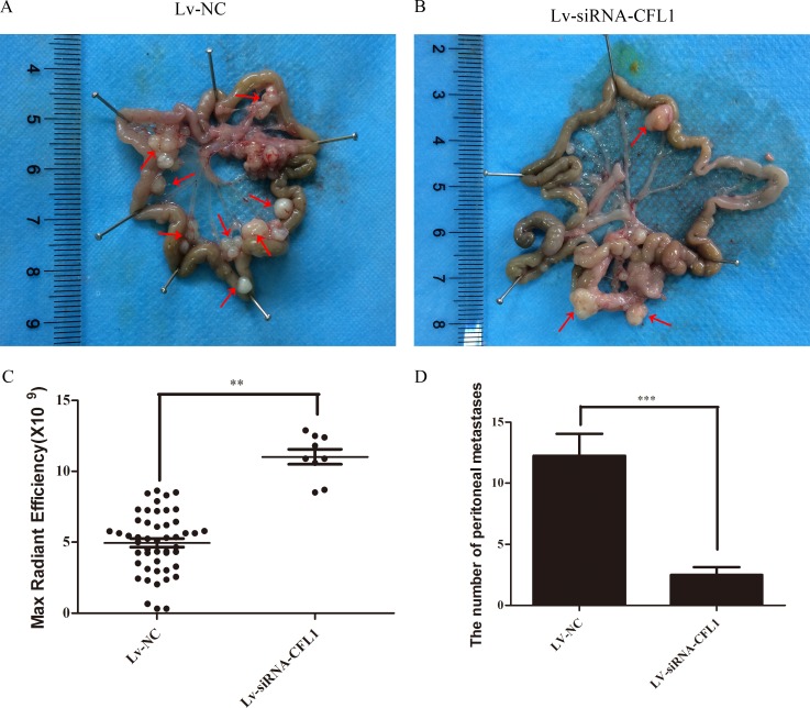
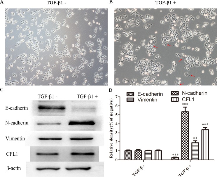
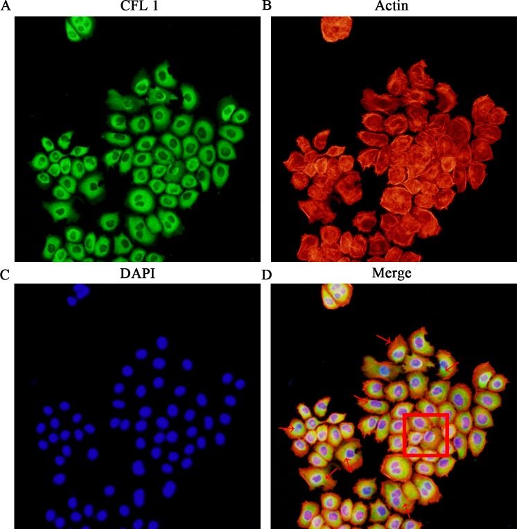
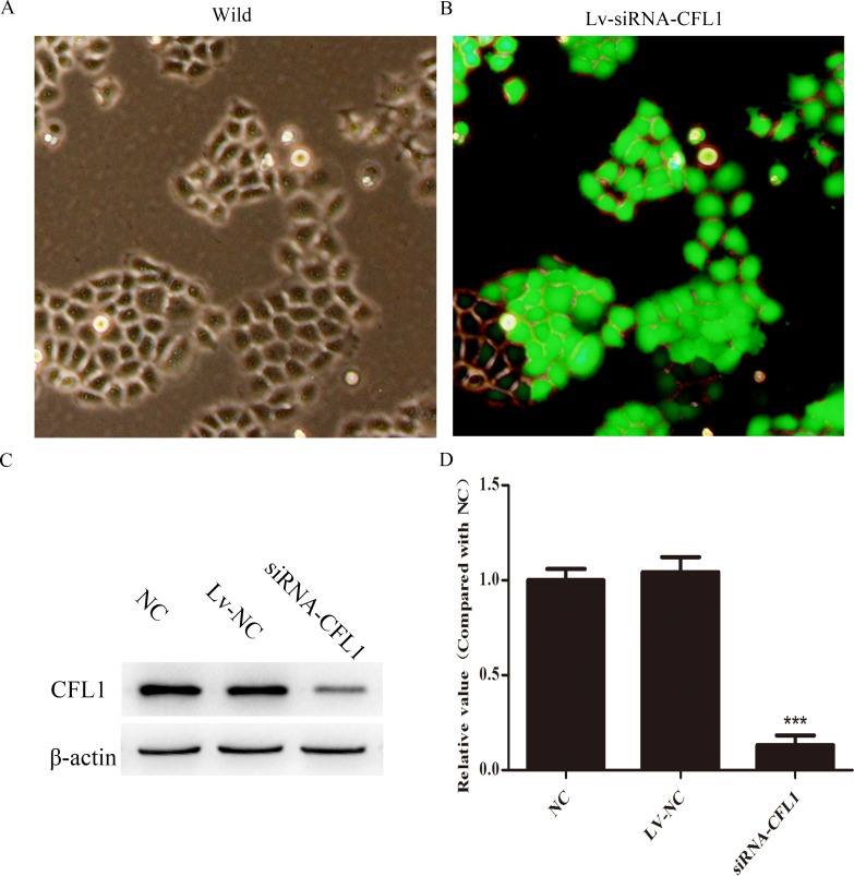
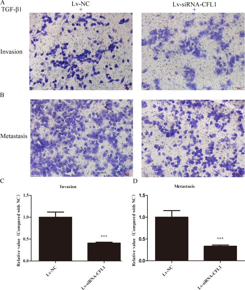
Epithelial-mesenchymal transition (EMT) is an important biological process whereby malignant tumor cells obtain the ability to migrate, invade, resist apoptosis and degrade the extracellular matrix. We found that Cofilin1 (CFL1) expression was elevated in clinical gastric cancer specimens and correlated with biomarkers of EMT in BGC-823 gastric cancer cells. BGC-823 cells exhibited EMT phenotypes and increased metastatic ability when induced by TGF-β1. By contrast, BGC-823 cells transfected with Lv-siRNA-CFL1 did not exhibit EMT phenotypes under the same inducing conditions. As CFL1 expression increased, EMT cell filopodia stretched out. In addition, the ultrastructures observed using transmission electron microscopy indicated that silencing of CFL1 markedly inhibited depolymerization of fibrous actin and cytoskeletal reorganization during EMT. Similar results were obtained in vivo. These findings demonstrate that CFL1 induces EMT by promoting cytoskeletal rearrangement. Our results may provide the basis for developing new anticancer drugs to inhibit CFL1.
上皮-间质转化(EMT)是一个重要的生物学过程,通过该过程恶性肿瘤细胞获得迁移、侵袭、抵抗凋亡和降解细胞外基质的能力。我们发现,Cofilin1(CFL1)在临床胃癌标本中的表达升高,且与BGC-823胃癌细胞中EMT的生物标志物相关。当受到转化生长因子-β1(TGF-β1)诱导时,BGC-823细胞表现出EMT表型且转移能力增强。相比之下,用Lv-siRNA-CFL1转染的BGC-823细胞在相同诱导条件下未表现出EMT表型。随着CFL1表达增加,EMT细胞丝状伪足伸展。此外,透射电子显微镜观察到的超微结构表明,CFL1的沉默显著抑制了EMT过程中纤维状肌动蛋白的解聚和细胞骨架重组。在体内也获得了类似结果。这些发现表明,CFL1通过促进细胞骨架重排诱导EMT。我们的结果可能为开发抑制CFL1的新型抗癌药物提供依据。