Mori Yuki, Murakami Tomoaki, Suzuma Kiyoshi, Ishihara Kenji, Yoshitake Shin, Fujimoto Masahiro, Dodo Yoko, Yoshitake Tatsuya, Miwa Yuko, Tsujikawa Akitaka
Department of Ophthalmology and Visual Sciences, Kyoto University Graduate School of Medicine, Kyoto, Japan.
PLoS One. 2017 Apr 13;12(4):e0175809. doi: 10.1371/journal.pone.0175809. eCollection 2017.
To investigate whether baseline optical coherence tomography (OCT) parameters can predict the treatment frequency of intravitreal ranibizumab (IVR) injections during the first year in patients with diabetic macular edema (DME) treated with pro re nata (PRN) IVR injections.
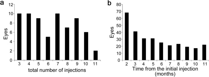
We retrospectively reviewed 68 eyes of 63 patients with center-involved DME who received IVR injections for 12 months or longer according to three monthly IVR injections followed by the PRN dosing. We measured the mean retinal thicknesses in the individual subfields of the Early Treatment Diabetic Retinopathy Study grid and evaluated the qualitative and quantitative parameters on OCT sectional images. We investigated the relationship between these OCT parameters at baseline and the number of IVR injections during the 12-month follow-up.
Three loading doses were administered to 10 eyes; four to seven annualized IVR injections were administered to 34 eyes. The number of eyes that received IVR injections decreased gradually until month 6 and was almost constant from months 7 to 11. No relationships were seen between the treatment frequency and baseline systemic factors and the ophthalmic examination findings. Univariate analyses showed that the number of IVR injections during the first year was associated with the mean retinal thickness in the individual subfields and the transverse length of the disrupted external limiting membrane (ELM) and ellipsoid zone of the photoreceptors. Multivariate analysis showed a significant association with the thickness in the inferior subfield alone. The treatment frequency during the 12-month follow-up was not correlated with improved visual acuity but was associated with the decrease in the central subfield thickness and disrupted ELM.
The retinal thickness in the inferior subfield predicts the treatment frequency during the first year in eyes with DME treated with PRN IVR injections.
探讨在接受按需玻璃体内注射雷珠单抗(IVR)治疗的糖尿病性黄斑水肿(DME)患者中,基线光学相干断层扫描(OCT)参数是否能够预测第一年的IVR注射治疗频率。
我们回顾性分析了63例累及黄斑中心凹的DME患者的68只眼,这些患者按照每月3次IVR注射后按需给药的方案接受了12个月或更长时间的IVR注射。我们测量了早期糖尿病性视网膜病变研究网格各个子区域的平均视网膜厚度,并评估了OCT断层图像上的定性和定量参数。我们研究了这些基线OCT参数与12个月随访期间IVR注射次数之间的关系。
10只眼接受了3次负荷剂量注射;34只眼每年接受4至7次IVR注射。接受IVR注射的眼数在第6个月前逐渐减少,在第7至11个月几乎保持不变。未发现治疗频率与基线全身因素及眼科检查结果之间存在关联。单因素分析显示,第一年的IVR注射次数与各个子区域的平均视网膜厚度以及光感受器外部限制膜(ELM)和椭圆体带的横向长度有关。多因素分析显示仅与下方子区域的厚度存在显著关联。12个月随访期间的治疗频率与视力改善无关,但与中心子区域厚度的降低和ELM的破坏有关。
下方子区域的视网膜厚度可预测按需IVR注射治疗的DME患眼第一年的治疗频率。