Caire Nail Laura, Rodríguez Reimundes Ezequiel, Weibel Galluzzo Christelle, Lebowitz Dan, Ibrahim Yasmine Lucile, Lobrinus Johannes Alexander, Chappuis François
Service de médecine interne générale, Hôpitaux Universitaires de Genève, Rue Gabrielle-Perret-Gentil 4, 1205, Geneva, Switzerland.
Service de médecine tropicale et humanitaire, Hôpitaux Universitaires de Genève, Rue Gabrielle-Perret-Gentil 4, 1205, Geneva, Switzerland.
J Med Case Rep. 2017 Apr 18;11(1):113. doi: 10.1186/s13256-017-1279-2.
Alveolar echinococcosis is a potentially lethal zoonosis caused by larval forms of the tapeworm Echinococcus multilocularis. Humans are aberrant intermediate hosts who become infected by ingestion of egg-contaminated food or water or via physical contact with domestic or wild animals that carry the parasite in their small intestine. In humans, the disease usually affects the liver and can spread to other organs causing metastatic infiltration. In this report, we describe an advanced presentation of human alveolar echinococcosis mimicking metastatic malignancy.
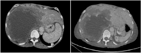
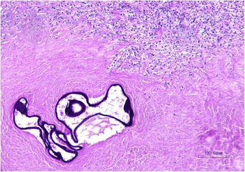
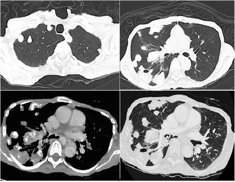
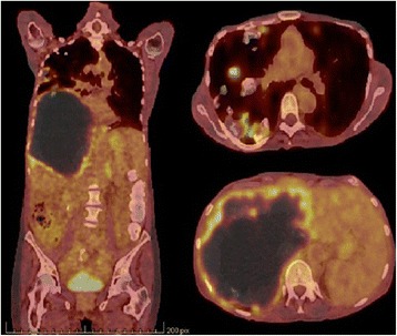
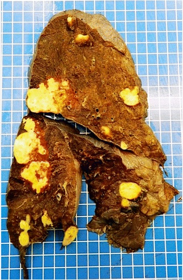
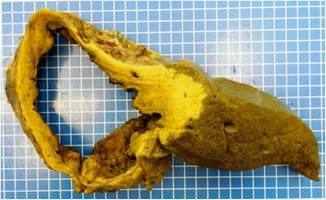
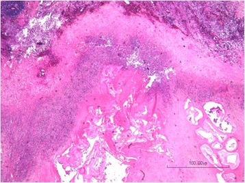
A 62-year-old white woman was evaluated for fever, jaundice, and abdominal pain, associated with significant weight loss. She lived in a rural area in Switzerland and used to eat wild forest fruits and mushrooms. She owned cats that used to hunt rodents. On physical examination, she appeared severely ill with cachexia, altered mental status, jaundice, and massive hepatomegaly. Laboratory tests showed cholestasis with preserved liver function. An abdominal computed tomography scan showed an enlarged liver with a huge cystic mass in the right lobe extending into the left lobe, infiltrating her hepatic hilum, causing intrahepatic bile duct dilation and occlusion of her right portal vein. A chest computed tomography scan showed multiple calcified bilateral pulmonary nodules. Her clinical and radiological presentation resembled an advanced neoplastic disease. Serologic tests for Echinococcus multilocularis were positive. The diagnosis of alveolar echinococcosis was established on her past history of exposure, imaging, and serology results.
Clinical presentation and radiologic imaging findings of disseminated alveolar echinococcosis can mimic metastatic malignancy, and diagnosis can be challenging in atypically advanced cases. As the incidence of human alveolar echinococcosis appears to be increasing in Europe and Switzerland, physicians should be aware of alveolar echinococcosis, its epidemiology, and its clinical features.
肺泡型棘球蚴病是一种由多房棘球绦虫幼虫引起的潜在致命性人畜共患病。人类是异常中间宿主,通过摄入受虫卵污染的食物或水,或通过与在小肠中携带该寄生虫的家畜或野生动物进行身体接触而感染。在人类中,该病通常影响肝脏,并可扩散至其他器官导致转移性浸润。在本报告中,我们描述了一例酷似转移性恶性肿瘤的人类肺泡型棘球蚴病的晚期表现。
一名62岁白人女性因发热、黄疸和腹痛就诊,伴有显著体重减轻。她生活在瑞士农村地区,过去常食用野生森林水果和蘑菇。她养了一些猫,这些猫过去常捕食啮齿动物。体格检查时,她表现出严重病态,伴有恶病质、精神状态改变、黄疸和肝脏肿大。实验室检查显示胆汁淤积,但肝功能正常。腹部计算机断层扫描显示肝脏肿大,右叶有一个巨大的囊性肿块延伸至左叶,侵犯肝门,导致肝内胆管扩张和右门静脉闭塞。胸部计算机断层扫描显示双侧多个钙化肺结节。她的临床和影像学表现类似于晚期肿瘤疾病。多房棘球绦虫的血清学检测呈阳性。根据她过去的接触史、影像学和血清学结果,确诊为肺泡型棘球蚴病。
播散性肺泡型棘球蚴病的临床表现和影像学检查结果可酷似转移性恶性肿瘤,在非典型晚期病例中诊断可能具有挑战性。由于欧洲和瑞士人类肺泡型棘球蚴病的发病率似乎在上升,医生应了解肺泡型棘球蚴病、其流行病学及其临床特征。