Lee Myungeun, Woo Boyeong, Kuo Michael D, Jamshidi Neema, Kim Jong Hyo
Center for Medical-IT Convergence Technology Research, Advanced Institutes of Convergence Technology, Seoul National University, Suwon 16229, Korea.
Department of Radiology, Seoul National University Hospital, Seoul 03080, Korea.
Korean J Radiol. 2017 May-Jun;18(3):498-509. doi: 10.3348/kjr.2017.18.3.498. Epub 2017 Apr 3.
The purpose of this study was to evaluate the reliability and quality of radiomic features in glioblastoma multiforme (GBM) derived from tumor volumes obtained with semi-automated tumor segmentation software.
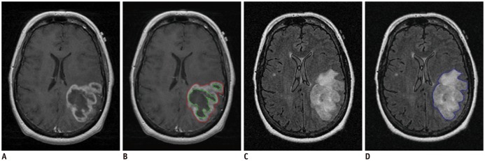
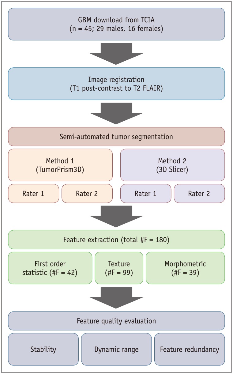
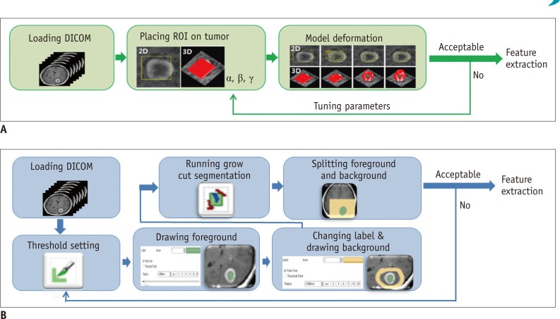
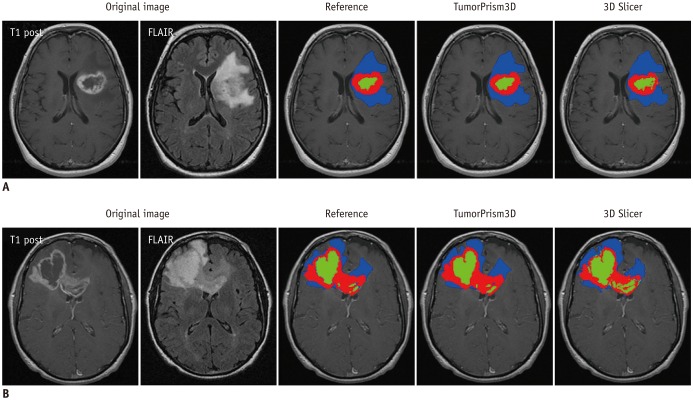
MR images of 45 GBM patients (29 males, 16 females) were downloaded from The Cancer Imaging Archive, in which post-contrast T1-weighted imaging and fluid-attenuated inversion recovery MR sequences were used. Two raters independently segmented the tumors using two semi-automated segmentation tools (TumorPrism3D and 3D Slicer). Regions of interest corresponding to contrast-enhancing lesion, necrotic portions, and non-enhancing T2 high signal intensity component were segmented for each tumor. A total of 180 imaging features were extracted, and their quality was evaluated in terms of stability, normalized dynamic range (NDR), and redundancy, using intra-class correlation coefficients, cluster consensus, and Rand Statistic.
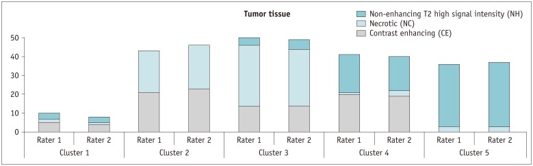
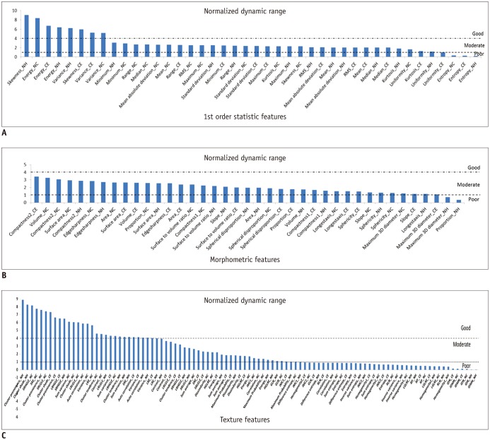
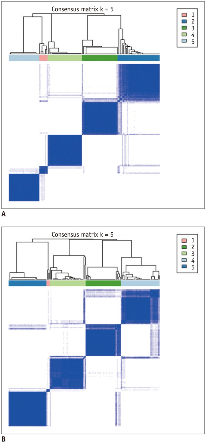
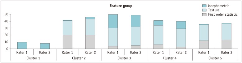
Our study results showed that most of the radiomic features in GBM were highly stable. Over 90% of 180 features showed good stability (intra-class correlation coefficient [ICC] ≥ 0.8), whereas only 7 features were of poor stability (ICC < 0.5). Most first order statistics and morphometric features showed moderate-to-high NDR (4 > NDR ≥1), while above 35% of the texture features showed poor NDR (< 1). Features were shown to cluster into only 5 groups, indicating that they were highly redundant.
The use of semi-automated software tools provided sufficiently reliable tumor segmentation and feature stability; thus helping to overcome the inherent inter-rater and intra-rater variability of user intervention. However, certain aspects of feature quality, including NDR and redundancy, need to be assessed for determination of representative signature features before further development of radiomics.
本研究旨在评估通过半自动肿瘤分割软件获得的多形性胶质母细胞瘤(GBM)肿瘤体积所衍生的放射组学特征的可靠性和质量。
从癌症影像存档库下载了45例GBM患者(29例男性,16例女性)的磁共振成像(MR)图像,其中使用了对比增强T1加权成像和液体衰减反转恢复MR序列。两名评估者使用两种半自动分割工具(TumorPrism3D和3D Slicer)独立分割肿瘤。为每个肿瘤分割出与对比增强病变、坏死部分和非增强T2高信号强度成分相对应的感兴趣区域。共提取了180个影像特征,并使用组内相关系数、聚类一致性和兰德统计量,从稳定性、归一化动态范围(NDR)和冗余性方面评估其质量。
我们的研究结果表明,GBM中的大多数放射组学特征高度稳定。180个特征中超过90%显示出良好的稳定性(组内相关系数[ICC]≥0.8),而只有7个特征稳定性较差(ICC<0.5)。大多数一阶统计量和形态计量学特征显示出中等到高的NDR(4>NDR≥1),而超过35%的纹理特征显示出较差的NDR(<1)。特征仅聚为5组,表明它们具有高度冗余性。
使用半自动软件工具可提供足够可靠的肿瘤分割和特征稳定性;从而有助于克服用户干预固有的评估者间和评估者内变异性。然而,在放射组学进一步发展之前,需要评估特征质量的某些方面,包括NDR和冗余性,以确定代表性的特征标记。