Department of Anatomy and Cell Biology, McGill University, Montréal, Québec, Canada H3A 0C7.
Structural and Computational Biology Unit, European Molecular Biology Laboratory, Heidelberg 69117, Germany.
Nat Commun. 2017 May 2;8:15035. doi: 10.1038/ncomms15035.
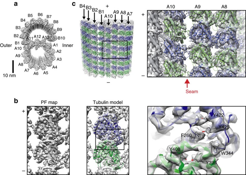
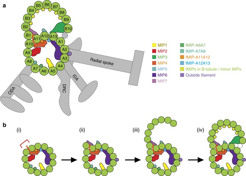
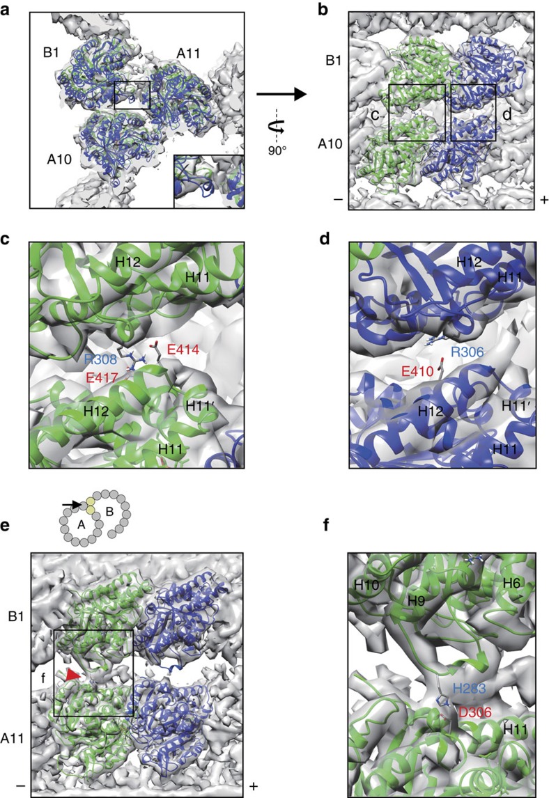
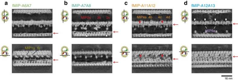
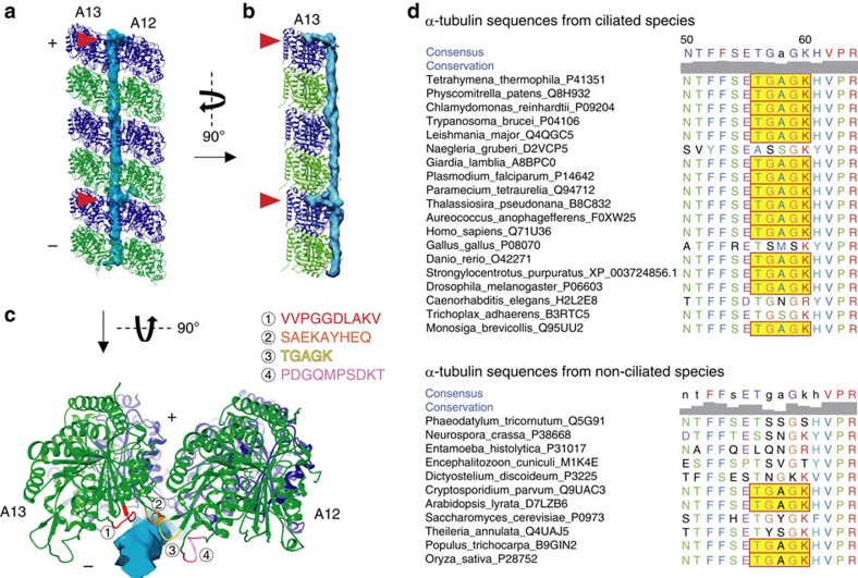
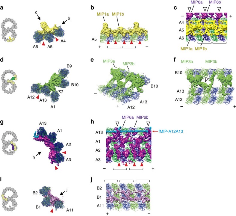
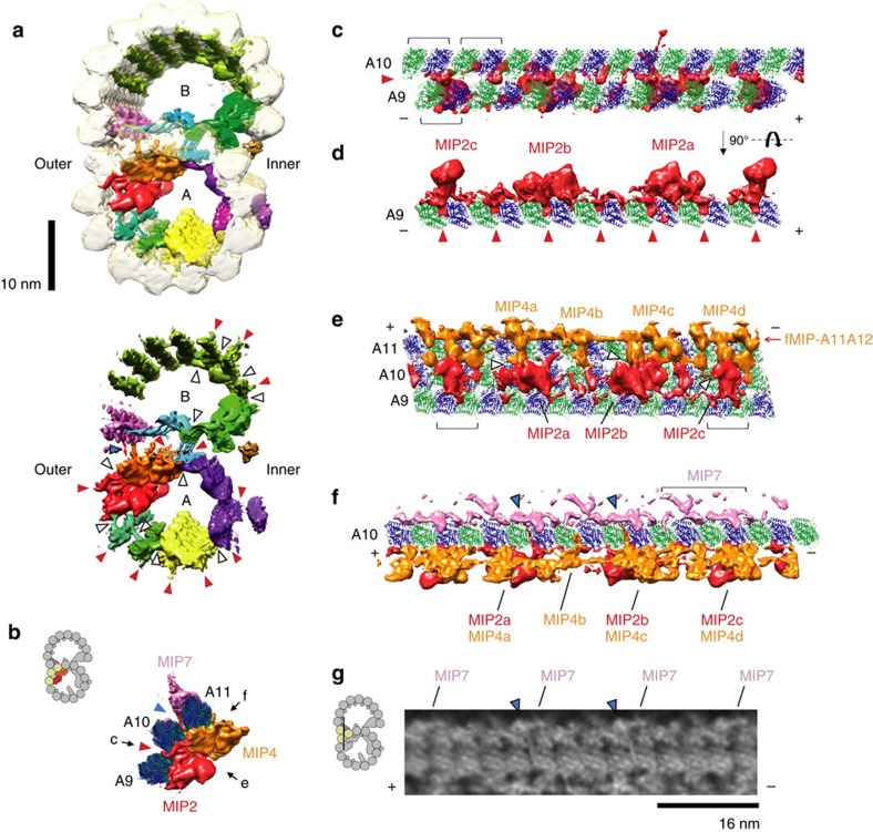
Cilia are ubiquitous, hair-like appendages found in eukaryotic cells that carry out functions of cell motility and sensory reception. Cilia contain an intriguing cytoskeletal structure, termed the axoneme that consists of nine doublet microtubules radially interlinked and longitudinally organized in multiple specific repeat units. Little is known, however, about how the axoneme allows cilia to be both actively bendable and sturdy or how it is assembled. To answer these questions, we used cryo-electron microscopy to structurally analyse several of the repeating units of the doublet at sub-nanometre resolution. This structural detail enables us to unambiguously assign α- and β-tubulins in the doublet microtubule lattice. Our study demonstrates the existence of an inner sheath composed of different kinds of microtubule inner proteins inside the doublet that likely stabilizes the structure and facilitates the specific building of the B-tubule.
纤毛是普遍存在于真核细胞中的毛发状附属物,具有细胞运动和感觉接收功能。纤毛含有一种有趣的细胞骨架结构,称为轴丝,由九个双联微管径向连接,并在多个特定重复单元中纵向排列。然而,人们对轴丝如何使纤毛既能灵活弯曲又坚固,以及它是如何组装的知之甚少。为了回答这些问题,我们使用低温电子显微镜以亚纳米分辨率对双联的几个重复单元进行结构分析。这种结构细节使我们能够在双联微管晶格中明确地分配α-和β-微管蛋白。我们的研究表明,在双联中存在一种由不同类型的微管内蛋白组成的内鞘,它可能稳定结构并促进 B-微管的特异性构建。