• 文献检索
  • 文档翻译
  • 深度研究
  • 学术资讯
  • Suppr Zotero 插件Zotero 插件
  • 邀请有礼
  • 套餐&价格
  • 历史记录
应用&插件
Suppr Zotero 插件Zotero 插件浏览器插件Mac 客户端Windows 客户端微信小程序
定价
高级版会员购买积分包购买API积分包
服务
文献检索文档翻译深度研究API 文档MCP 服务
关于我们
关于 Suppr公司介绍联系我们用户协议隐私条款
关注我们

Suppr 超能文献

核心技术专利:CN118964589B侵权必究
粤ICP备2023148730 号-1Suppr @ 2026

文献检索

告别复杂PubMed语法,用中文像聊天一样搜索,搜遍4000万医学文献。AI智能推荐,让科研检索更轻松。

立即免费搜索

文件翻译

保留排版,准确专业,支持PDF/Word/PPT等文件格式,支持 12+语言互译。

免费翻译文档

深度研究

AI帮你快速写综述,25分钟生成高质量综述,智能提取关键信息,辅助科研写作。

立即免费体验

通过荧光光学成像检测系统性硬化症患者手部微循环紊乱:一项初步研究。

Disturbed microcirculation in the hands of patients with systemic sclerosis detected by fluorescence optical imaging: a pilot study.

作者信息

Friedrich Stefanie, Lüders Susanne, Werner Stephanie Gabriele, Glimm Anne-Marie, Burmester Gerd-Rüdiger, Riemekasten Gabriela, Backhaus Marina, Ohrndorf Sarah

机构信息

Department of Rheumatology and Clinical Immunology, Charité University Hospital, Berlin, Germany.

Rheumatologie, Immunologie und Osteologie mit, Schwerpunkt für Rheumatologie, Klinische Immunologie und Osteologie am Ev. Krankenhaus Düsseldorf, Düsseldorf, Germany.

出版信息

Arthritis Res Ther. 2017 May 8;19(1):87. doi: 10.1186/s13075-017-1300-6.

DOI:10.1186/s13075-017-1300-6
PMID:28482872
原文链接:https://pmc.ncbi.nlm.nih.gov/articles/PMC5422953/
Abstract

BACKGROUND

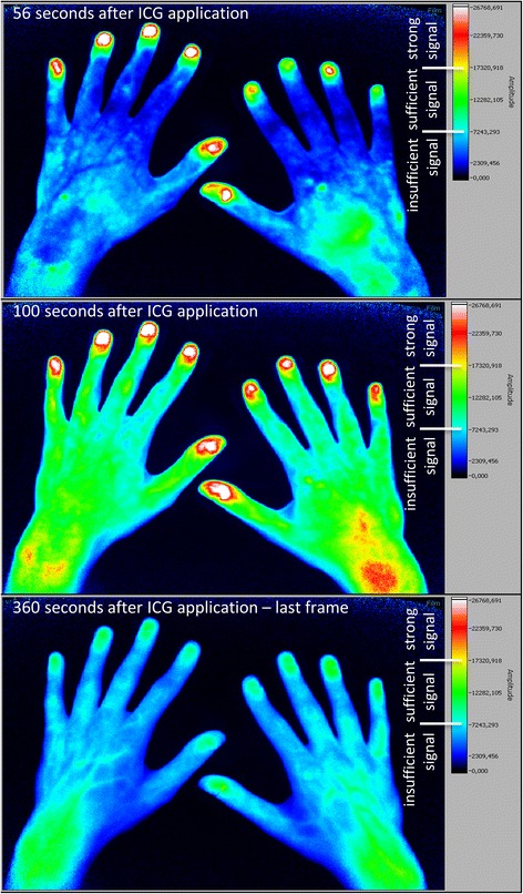
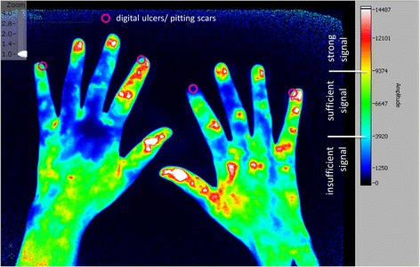
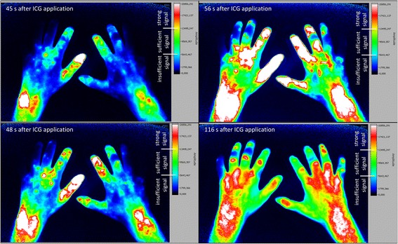
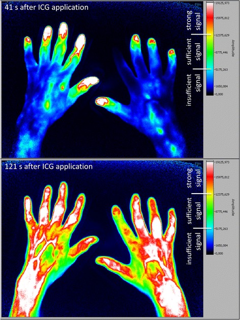
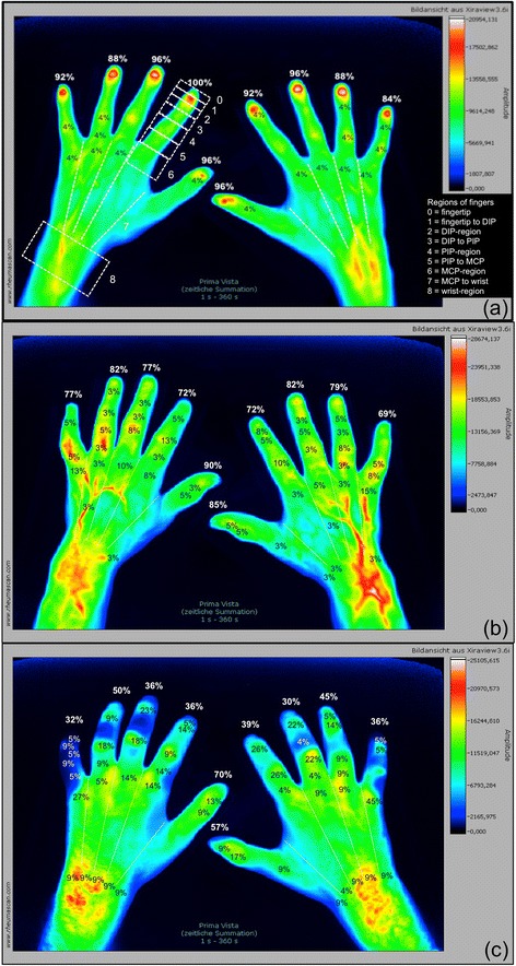
Utilising fluorescence optical imaging (FOI), the distribution of an intravenously applied colouring agent indocyanine green (ICG) can be analysed with the potential to identify malperfusion by little to no tissue enhancement. Systemic sclerosis (SSc) is characterised by the presence of digital ulcers reflecting progressive vasculopathy. The objective was to investigate the potential of FOI in the detection of disturbed microcirculation in the hands and fingers of patients with SSc and to link FOI findings to clinical signs of ischemia such as digital ulcers and pitting scars.

METHODS

In this cross-sectional study, 63 patients with SSc and 26 healthy subjects were examined. FOI was performed in all 89 individuals and compared to clinical data and capillaroscopic findings assembled for the SSc cohort.

RESULTS

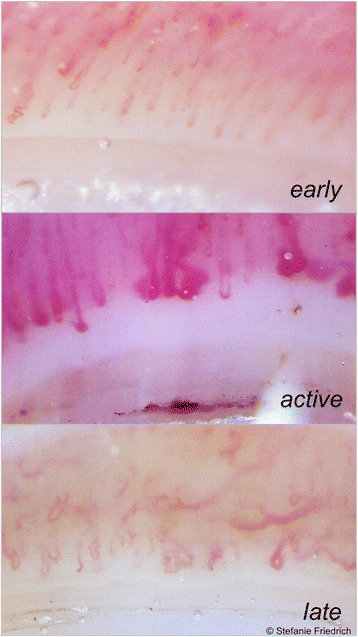
Healthy subjects showed initial ICG signals in their fingertips in 93.6%, SSc patients in 78.5% (limited SSc) and 43.2% (diffuse SSc). Moreover, in SSc patients, FOI findings were significantly associated with a late capillaroscopic pattern, disseminated SSc features, a diffuse SSc subtype, and the presence of digital ulcers or pitting scars. Intra- and inter-reader reliability for FOI amounted to κ = 0.786 and κ = 0.834, respectively.

CONCLUSIONS

FOI is able to detect areas of reduced microcirculation in patients with SSc with high association to capillaroscopic findings. The results pave the way for future FOI investigations into its role in the prediction of complications due to an impaired acral perfusion.

摘要

背景

利用荧光光学成像(FOI),可以分析静脉注射的染色剂吲哚菁绿(ICG)的分布情况,其有潜力通过极少或无组织增强来识别灌注不良。系统性硬化症(SSc)的特征是存在反映进行性血管病变的指端溃疡。目的是研究FOI在检测SSc患者手部和手指微循环紊乱方面的潜力,并将FOI结果与缺血的临床体征(如指端溃疡和点状瘢痕)联系起来。

方法

在这项横断面研究中,对63例SSc患者和26名健康受试者进行了检查。对所有89名个体进行了FOI检查,并与为SSc队列收集的临床数据和毛细血管镜检查结果进行了比较。

结果

93.6%的健康受试者指尖出现初始ICG信号,局限性SSc患者中这一比例为78.5%,弥漫性SSc患者中为43.2%。此外,在SSc患者中,FOI结果与晚期毛细血管镜检查模式、弥漫性SSc特征、弥漫性SSc亚型以及指端溃疡或点状瘢痕的存在显著相关。FOI的阅片者内和阅片者间可靠性分别为κ = 0.786和κ = 0.834。

结论

FOI能够检测出SSc患者微循环减少的区域,且与毛细血管镜检查结果高度相关。这些结果为未来关于FOI在预测因肢端灌注受损导致的并发症中作用的研究铺平了道路。

https://cdn.ncbi.nlm.nih.gov/pmc/blobs/c272/5422953/b315eff6171e/13075_2017_1300_Fig6_HTML.jpg
https://cdn.ncbi.nlm.nih.gov/pmc/blobs/c272/5422953/f6fb8cf872e9/13075_2017_1300_Fig1_HTML.jpg
https://cdn.ncbi.nlm.nih.gov/pmc/blobs/c272/5422953/150974758ee8/13075_2017_1300_Fig2_HTML.jpg
https://cdn.ncbi.nlm.nih.gov/pmc/blobs/c272/5422953/4f25e7eb6404/13075_2017_1300_Fig3_HTML.jpg
https://cdn.ncbi.nlm.nih.gov/pmc/blobs/c272/5422953/6f94a3963503/13075_2017_1300_Fig4_HTML.jpg
https://cdn.ncbi.nlm.nih.gov/pmc/blobs/c272/5422953/07ed85e684ae/13075_2017_1300_Fig5_HTML.jpg
https://cdn.ncbi.nlm.nih.gov/pmc/blobs/c272/5422953/b315eff6171e/13075_2017_1300_Fig6_HTML.jpg

相似文献

1
Disturbed microcirculation in the hands of patients with systemic sclerosis detected by fluorescence optical imaging: a pilot study.通过荧光光学成像检测系统性硬化症患者手部微循环紊乱:一项初步研究。
Arthritis Res Ther. 2017 May 8;19(1):87. doi: 10.1186/s13075-017-1300-6.
2
Association between baseline clinical and imaging findings and the development of digital ulcers in patients with systemic sclerosis.系统性硬化症患者的基线临床和影像学表现与发生手指溃疡的相关性。
Arthritis Res Ther. 2019 Apr 15;21(1):96. doi: 10.1186/s13075-019-1875-1.
3
The first composite score predicting Digital Ulcers in systemic sclerosis patients using Clinical data, Imaging and Patient history-CIP-DUS.使用临床数据、影像学和患者病史预测系统性硬化症患者出现 Digital Ulcers 的首个综合评分-CIP-DUS。
Arthritis Res Ther. 2020 Jun 15;22(1):144. doi: 10.1186/s13075-020-02235-7.
4
Comparison of laser Doppler imaging, fingertip lacticemy test, and nailfold capillaroscopy for assessment of digital microcirculation in systemic sclerosis.激光多普勒成像、指尖乳酸试验和甲襞毛细血管镜检查评估系统性硬化症手指微循环的比较。
Arthritis Res Ther. 2010;12(4):R157. doi: 10.1186/ar3112. Epub 2010 Aug 10.
5
The Application of Fluorescence Optical Imaging in Systemic Sclerosis.荧光光学成像在系统性硬化症中的应用
Biomed Res Int. 2015;2015:658710. doi: 10.1155/2015/658710. Epub 2015 Aug 18.
6
Capillaroscopic findings in systemic sclerosis -- are they associated with disease duration and presence of digital ulcers?系统性硬化症的毛细血管镜检查结果——它们与疾病持续时间和指端溃疡的存在有关吗?
Discov Med. 2011 Nov;12(66):413-8.
7
Capillaroscopic pattern at the toes of systemic sclerosis patients: does it "tell" more than those of fingers?系统性硬化症患者脚趾的毛细血管镜模式:它比手指的“告诉”更多吗?
J Clin Rheumatol. 2011 Sep;17(6):311-4. doi: 10.1097/RHU.0b013e31822be4e8.
8
The association between endothelial microparticles and inflammation in patients with systemic sclerosis and Raynaud's phenomenon as detected by functional imaging.通过功能成像检测系统性硬化症和雷诺现象患者内皮微粒与炎症之间的关联。
Clin Hemorheol Microcirc. 2015;61(3):549-57. doi: 10.3233/CH-151956.
9
Inhomogeneity of capillaroscopic findings in systemic sclerosis.系统性硬化症中毛细血管镜检查结果的不均匀性。
Int J Rheum Dis. 2020 Feb;23(2):207-215. doi: 10.1111/1756-185X.13760. Epub 2019 Dec 5.
10
Abnormal amplitude and kinetics of digital postocclusive reactive hyperemia in systemic sclerosis.系统性硬化症中手指阻断后反应性充血的异常幅度和动力学
Microvasc Res. 2014 Jul;94:90-5. doi: 10.1016/j.mvr.2014.05.007. Epub 2014 Jun 2.

引用本文的文献

1
To optimise the diagnostic process of rheumatic diseases affecting the hands using fluorescence optical imaging (FOI).利用荧光光学成像(FOI)优化手部风湿性疾病的诊断流程。
RMD Open. 2025 Aug 31;11(3):e005372. doi: 10.1136/rmdopen-2024-005372.
2
Botulinum Toxin for the Treatment of Raynaud's Conditions of the Hand: Clinical Practice Updates and Future Directions.肉毒毒素治疗手部雷诺现象:临床实践更新及未来方向。
Toxins (Basel). 2024 Nov 1;16(11):472. doi: 10.3390/toxins16110472.
3
Impact of light polarization on laser speckle contrast imaging with a custom phantom for microvascular flow.

本文引用的文献

1
Detection of severe digital vasculopathy in systemic sclerosis by colour Doppler sonography is associated with digital ulcers.通过彩色多普勒超声检测系统性硬化症中的严重指端血管病变与指端溃疡相关。
Rheumatology (Oxford). 2017 Nov 1;56(11):1865-1873. doi: 10.1093/rheumatology/kex045.
2
Capillaroscopy 2016: new perspectives in systemic sclerosis.2016年毛细血管显微镜检查:系统性硬化症的新视角
Acta Reumatol Port. 2016 Jan-Mar;41(1):8-14.
3
Nailfold Videocapillaroscopic Features and Other Clinical Risk Factors for Digital Ulcers in Systemic Sclerosis: A Multicenter, Prospective Cohort Study.
光偏振对微血管流动定制体模激光散斑对比成像的影响。
Sci Rep. 2024 Nov 4;14(1):26652. doi: 10.1038/s41598-024-73757-2.
4
Metabolic and molecular imaging in inflammatory arthritis.炎症性关节炎的代谢与分子成像
RMD Open. 2024 Feb 9;10(1):e003880. doi: 10.1136/rmdopen-2023-003880.
5
Fluorescence optical imaging feature selection with machine learning for differential diagnosis of selected rheumatic diseases.基于机器学习的荧光光学成像特征选择用于特定风湿性疾病的鉴别诊断
Front Med (Lausanne). 2023 Aug 21;10:1228833. doi: 10.3389/fmed.2023.1228833. eCollection 2023.
6
[Can rheuma be scanned? : Review of the current study situation on fluorescence optical imaging].[类风湿病可以进行扫描吗?:荧光光学成像当前研究状况综述]
Z Rheumatol. 2023 Oct;82(8):627-637. doi: 10.1007/s00393-023-01404-8. Epub 2023 Aug 25.
7
Features Found in Indocyanine Green-Based Fluorescence Optical Imaging of Inflammatory Diseases of the Hands.基于吲哚菁绿的手部炎症性疾病荧光光学成像中发现的特征。
Diagnostics (Basel). 2022 Jul 22;12(8):1775. doi: 10.3390/diagnostics12081775.
8
Detection of microvascular changes in systemic sclerosis and other rheumatic diseases.系统性硬化症和其他风湿性疾病的微血管变化检测。
Nat Rev Rheumatol. 2021 Nov;17(11):665-677. doi: 10.1038/s41584-021-00685-0. Epub 2021 Sep 24.
9
Fluorescence optical imaging: ready for prime time?荧光光学成像:是否已准备好进入黄金时期?
RMD Open. 2021 Jun;7(2). doi: 10.1136/rmdopen-2020-001497.
10
Detection of subclinical skin manifestation in patients with psoriasis and psoriatic arthritis by fluorescence optical imaging.通过荧光光学成像检测银屑病和银屑病关节炎患者的亚临床皮肤表现。
Arthritis Res Ther. 2020 Aug 18;22(1):192. doi: 10.1186/s13075-020-02277-x.
甲襞微循环视频镜检特征和系统性硬化症发生手指溃疡的其他临床危险因素:一项多中心前瞻性队列研究。
Arthritis Rheumatol. 2016 Oct;68(10):2527-39. doi: 10.1002/art.39718.
4
Nailfold Capillaroscopy and Clinical Applications in Systemic Sclerosis.甲襞毛细血管镜检查及其在系统性硬化症中的临床应用
Microcirculation. 2016 Jul;23(5):364-72. doi: 10.1111/micc.12281.
5
Reliability of the quantitative assessment of peripheral blood perfusion by laser speckle contrast analysis in a systemic sclerosis cohort.系统性硬化症队列中通过激光散斑对比分析对外周血灌注进行定量评估的可靠性
Ann Rheum Dis. 2016 Jun;75(6):1263-4. doi: 10.1136/annrheumdis-2015-208857. Epub 2016 Mar 22.
6
The Application of Fluorescence Optical Imaging in Systemic Sclerosis.荧光光学成像在系统性硬化症中的应用
Biomed Res Int. 2015;2015:658710. doi: 10.1155/2015/658710. Epub 2015 Aug 18.
7
Analysis of distribution and severity of inflammation in patients with osteoarthitis compared to rheumatoid arthritis by ICG-enhanced fluorescence optical imaging and musculoskeletal ultrasound: a pilot study.通过吲哚菁绿增强荧光光学成像和肌肉骨骼超声分析骨关节炎与类风湿关节炎患者炎症的分布及严重程度:一项初步研究。
Ann Rheum Dis. 2016 Mar;75(3):566-70. doi: 10.1136/annrheumdis-2015-207345. Epub 2015 Aug 26.
8
Short-term follow-up of digital ulcers by laser speckle contrast analysis in systemic sclerosis patients.系统性硬化症患者数字溃疡的激光散斑对比分析短期随访
Microvasc Res. 2015 Sep;101:82-5. doi: 10.1016/j.mvr.2015.06.009. Epub 2015 Jun 30.
9
Endothelial Dysfunction and Nailfold Videocapillaroscopy Pattern as Predictors of Digital Ulcers in Systemic Sclerosis: a Cohort Study and Review of the Literature.内皮功能障碍和甲襞微血管镜检查模式作为系统性硬化症中手指溃疡的预测指标:一项队列研究及文献综述
Clin Rev Allergy Immunol. 2015 Oct;49(2):240-52. doi: 10.1007/s12016-015-8500-0.
10
A PRISMA-driven systematic review for predictive risk factors of digital ulcers in systemic sclerosis patients.基于 PRISMA 的系统性硬化症患者发生手指溃疡预测性风险因素的系统评价。
Autoimmun Rev. 2015 Feb;14(2):140-52. doi: 10.1016/j.autrev.2014.10.009. Epub 2014 Oct 14.