• 文献检索
  • 文档翻译
  • 深度研究
  • 学术资讯
  • Suppr Zotero 插件Zotero 插件
  • 邀请有礼
  • 套餐&价格
  • 历史记录
应用&插件
Suppr Zotero 插件Zotero 插件浏览器插件Mac 客户端Windows 客户端微信小程序
定价
高级版会员购买积分包购买API积分包
服务
文献检索文档翻译深度研究API 文档MCP 服务
关于我们
关于 Suppr公司介绍联系我们用户协议隐私条款
关注我们

Suppr 超能文献

核心技术专利:CN118964589B侵权必究
粤ICP备2023148730 号-1Suppr @ 2026

文献检索

告别复杂PubMed语法,用中文像聊天一样搜索,搜遍4000万医学文献。AI智能推荐,让科研检索更轻松。

立即免费搜索

文件翻译

保留排版,准确专业,支持PDF/Word/PPT等文件格式,支持 12+语言互译。

免费翻译文档

深度研究

AI帮你快速写综述,25分钟生成高质量综述,智能提取关键信息,辅助科研写作。

立即免费体验

光谱成像工具箱:用于确定细胞和模型膜脂质有序性的光谱图像分割、超堆栈重建及批处理。

Spectral imaging toolbox: segmentation, hyperstack reconstruction, and batch processing of spectral images for the determination of cell and model membrane lipid order.

作者信息

Aron Miles, Browning Richard, Carugo Dario, Sezgin Erdinc, Bernardino de la Serna Jorge, Eggeling Christian, Stride Eleanor

机构信息

Department of Engineering Science, Institute of Biomedical Engineering, University of Oxford, Oxford, OX3 7DQ, UK.

Faculty of Engineering and The Environment, University of Southampton, Southampton, SO17 1BJ, UK.

出版信息

BMC Bioinformatics. 2017 May 12;18(1):254. doi: 10.1186/s12859-017-1656-2.

DOI:10.1186/s12859-017-1656-2
PMID:28494801
原文链接:https://pmc.ncbi.nlm.nih.gov/articles/PMC5427590/
Abstract

BACKGROUND

Spectral imaging with polarity-sensitive fluorescent probes enables the quantification of cell and model membrane physical properties, including local hydration, fluidity, and lateral lipid packing, usually characterized by the generalized polarization (GP) parameter. With the development of commercial microscopes equipped with spectral detectors, spectral imaging has become a convenient and powerful technique for measuring GP and other membrane properties. The existing tools for spectral image processing, however, are insufficient for processing the large data sets afforded by this technological advancement, and are unsuitable for processing images acquired with rapidly internalized fluorescent probes.

RESULTS

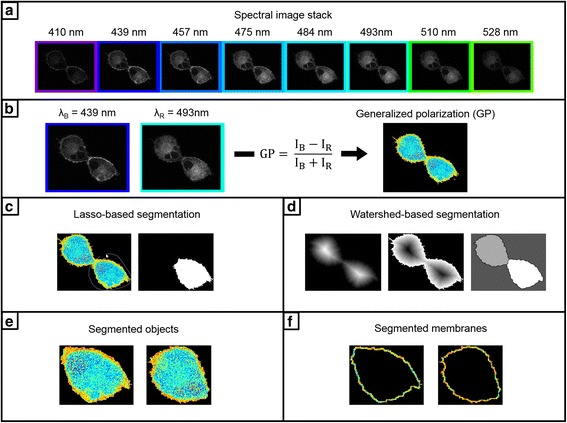
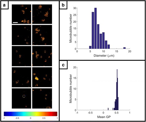
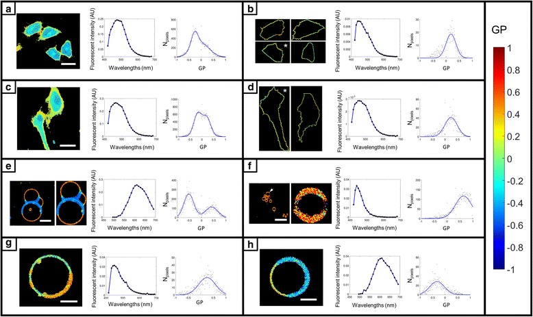
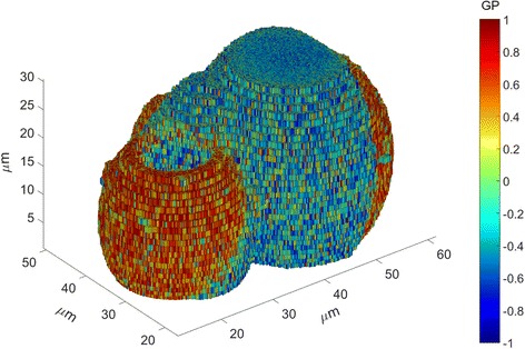
Here we present a MATLAB spectral imaging toolbox with the aim of overcoming these limitations. In addition to common operations, such as the calculation of distributions of GP values, generation of pseudo-colored GP maps, and spectral analysis, a key highlight of this tool is reliable membrane segmentation for probes that are rapidly internalized. Furthermore, handling for hyperstacks, 3D reconstruction and batch processing facilitates analysis of data sets generated by time series, z-stack, and area scan microscope operations. Finally, the object size distribution is determined, which can provide insight into the mechanisms underlying changes in membrane properties and is desirable for e.g. studies involving model membranes and surfactant coated particles. Analysis is demonstrated for cell membranes, cell-derived vesicles, model membranes, and microbubbles with environmentally-sensitive probes Laurdan, carboxyl-modified Laurdan (C-Laurdan), Di-4-ANEPPDHQ, and Di-4-AN(F)EPPTEA (FE), for quantification of the local lateral density of lipids or lipid packing.

CONCLUSIONS

The Spectral Imaging Toolbox is a powerful tool for the segmentation and processing of large spectral imaging datasets with a reliable method for membrane segmentation and no ability in programming required. The Spectral Imaging Toolbox can be downloaded from https://uk.mathworks.com/matlabcentral/fileexchange/62617-spectral-imaging-toolbox .

摘要

背景

使用极性敏感荧光探针进行光谱成像能够量化细胞和模型膜的物理性质,包括局部水合作用、流动性和横向脂质堆积,通常用广义极化(GP)参数来表征。随着配备光谱探测器的商用显微镜的发展,光谱成像已成为测量GP和其他膜性质的便捷且强大的技术。然而,现有的光谱图像处理工具不足以处理这项技术进步所带来的大量数据集,并且不适用于处理使用快速内化荧光探针获取的图像。

结果

在此,我们展示了一个MATLAB光谱成像工具箱,旨在克服这些限制。除了常见操作,如计算GP值分布、生成伪彩色GP图和光谱分析外,该工具的一个关键亮点是对快速内化探针进行可靠的膜分割。此外,对超堆栈的处理、三维重建和批处理有助于分析由时间序列、z堆栈和面积扫描显微镜操作生成的数据集。最后,确定对象大小分布,这可以深入了解膜性质变化的潜在机制,并且对于例如涉及模型膜和表面活性剂包被颗粒的研究很有必要。使用对环境敏感的探针Laurdan、羧基修饰的Laurdan(C-Laurdan)、Di-4-ANEPPDHQ和Di-4-AN(F)EPPTEA(FE)对细胞膜、细胞衍生囊泡、模型膜和微泡进行分析,以量化脂质的局部横向密度或脂质堆积。

结论

光谱成像工具箱是用于分割和处理大型光谱成像数据集的强大工具,具有可靠的膜分割方法且无需编程能力。光谱成像工具箱可从https://uk.mathworks.com/matlabcentral/fileexchange/62617-spectral-imaging-toolbox下载。

https://cdn.ncbi.nlm.nih.gov/pmc/blobs/cdd9/5427590/943964c15e37/12859_2017_1656_Fig4_HTML.jpg
https://cdn.ncbi.nlm.nih.gov/pmc/blobs/cdd9/5427590/78dbe684f1f4/12859_2017_1656_Fig1_HTML.jpg
https://cdn.ncbi.nlm.nih.gov/pmc/blobs/cdd9/5427590/b0c44a299ee9/12859_2017_1656_Fig2_HTML.jpg
https://cdn.ncbi.nlm.nih.gov/pmc/blobs/cdd9/5427590/043e222cc263/12859_2017_1656_Fig3_HTML.jpg
https://cdn.ncbi.nlm.nih.gov/pmc/blobs/cdd9/5427590/943964c15e37/12859_2017_1656_Fig4_HTML.jpg

相似文献

1
Spectral imaging toolbox: segmentation, hyperstack reconstruction, and batch processing of spectral images for the determination of cell and model membrane lipid order.光谱成像工具箱:用于确定细胞和模型膜脂质有序性的光谱图像分割、超堆栈重建及批处理。
BMC Bioinformatics. 2017 May 12;18(1):254. doi: 10.1186/s12859-017-1656-2.
2
The spectral phasor approach to resolving membrane order with environmentally sensitive dyes.用环境敏感染料解析膜有序性的光谱相位子方法。
Methods Enzymol. 2024;700:105-126. doi: 10.1016/bs.mie.2024.01.024. Epub 2024 Feb 5.
3
Disorder Amidst Membrane Order: Standardizing Laurdan Generalized Polarization and Membrane Fluidity Terms.膜有序中的无序:标准化劳丹各向异性参数和膜流动性术语。
J Fluoresc. 2017 Jan;27(1):243-249. doi: 10.1007/s10895-016-1951-8. Epub 2016 Oct 13.
4
Time-Resolved Laurdan Fluorescence Reveals Insights into Membrane Viscosity and Hydration Levels.时间分辨劳丹荧光法揭示了对膜粘度和水合水平的深入了解。
Biophys J. 2018 Oct 16;115(8):1498-1508. doi: 10.1016/j.bpj.2018.08.041. Epub 2018 Sep 6.
5
Laurdan and di-4-ANEPPDHQ do not respond to membrane-inserted peptides and are good probes for lipid packing.劳丹荧光染料和二-4-ANEPPDHQ对插入膜内的肽不产生反应,是用于脂质堆积研究的优良探针。
Biochim Biophys Acta. 2011 Jan;1808(1):298-306. doi: 10.1016/j.bbamem.2010.10.002. Epub 2010 Oct 16.
6
LAURDAN since Weber: The Quest for Visualizing Membrane Heterogeneity.从劳丹到韦伯:探索膜异质性的可视化。
Acc Chem Res. 2021 Feb 16;54(4):976-987. doi: 10.1021/acs.accounts.0c00687. Epub 2021 Jan 29.
7
Optimized time-gated generalized polarization imaging of Laurdan and di-4-ANEPPDHQ for membrane order image contrast enhancement.优化的时门控广义偏振成像 Laurdan 和二-4-ANEPPDHQ 用于增强膜有序度图像对比。
Microsc Res Tech. 2010 Jun;73(6):618-22. doi: 10.1002/jemt.20801.
8
Imaging lipid lateral organization in membranes with C-laurdan in a confocal microscope.在共聚焦显微镜中使用 C-胆甾醇在膜中成像脂质的层状组织。
J Lipid Res. 2012 Mar;53(3):609-616. doi: 10.1194/jlr.D021311. Epub 2011 Dec 19.
9
Melittin Induces Local Order Changes in Artificial and Biological Membranes as Revealed by Spectral Analysis of Laurdan Fluorescence.蜂毒素诱导人工和生物膜的局部有序变化,如通过劳丹荧光的光谱分析所揭示的。
Toxins (Basel). 2020 Nov 8;12(11):705. doi: 10.3390/toxins12110705.
10
Microfluorometry of cell membrane dynamics.细胞膜动力学的显微荧光测定法。
Cytometry A. 2006 Mar;69(3):185-8. doi: 10.1002/cyto.a.20233.

引用本文的文献

1
Measuring plasma membrane fluidity using confocal microscopy.使用共聚焦显微镜测量质膜流动性。
Nat Protoc. 2025 Feb 19. doi: 10.1038/s41596-024-01122-8.
2
VISION - an open-source software for automated multi-dimensional image analysis of cellular biophysics.VISION——一个用于细胞生物物理学的自动化多维图像分析的开源软件。
J Cell Sci. 2024 Oct 15;137(20). doi: 10.1242/jcs.262166. Epub 2024 Oct 23.
3
Biophysical Interaction Landscape of Mycobacterial Mycolic Acids and Phenolic Glycolipids with Host Macrophage Membranes.分枝杆菌的类脂阿拉伯甘露聚糖和酚类糖脂与宿主巨噬细胞膜的生物物理相互作用图谱。

本文引用的文献

1
Modulation of the molecular arrangement in artificial and biological membranes by phospholipid-shelled microbubbles.磷脂壳微泡对人工和生物膜中分子排列的调节。
Biomaterials. 2017 Jan;113:105-117. doi: 10.1016/j.biomaterials.2016.10.034. Epub 2016 Oct 24.
2
The ImageJ ecosystem: An open platform for biomedical image analysis.ImageJ生态系统:一个用于生物医学图像分析的开放平台。
Mol Reprod Dev. 2015 Jul-Aug;82(7-8):518-29. doi: 10.1002/mrd.22489. Epub 2015 Jul 7.
3
Biologically and Acoustically Compatible Chamber for Studying Ultrasound-Mediated Delivery of Therapeutic Compounds.
ACS Appl Bio Mater. 2023 Dec 18;6(12):5555-5562. doi: 10.1021/acsabm.3c00748. Epub 2023 Nov 28.
4
Both Clathrin-Mediated and Membrane Microdomain-Associated Endocytosis Contribute to the Cellular Adaptation to Hyperosmotic Stress in .网格蛋白介导的内吞作用和膜微区相关的内吞作用均有助于 细胞适应高渗应激。
Int J Mol Sci. 2021 Nov 21;22(22):12534. doi: 10.3390/ijms222212534.
5
Monoethanolamine-induced glucose deprivation promotes apoptosis through metabolic rewiring in prostate cancer.单乙醇胺诱导的葡萄糖剥夺通过代谢重排促进前列腺癌细胞凋亡。
Theranostics. 2021 Aug 27;11(18):9089-9106. doi: 10.7150/thno.62724. eCollection 2021.
6
Toward Precisely Controllable Acoustic Response of Shell-Stabilized Nanobubbles: High Yield and Narrow Dispersity.实现壳层稳定纳米气泡精确可控的声学响应:高产率与窄分散性
ACS Nano. 2021 Mar 23;15(3):4901-4915. doi: 10.1021/acsnano.0c09701. Epub 2021 Mar 8.
7
Patches and Blebs: A Comparative Study of the Composition and Biophysical Properties of Two Plasma Membrane Preparations from CHO Cells.斑块和疱:两种 CHO 细胞膜制剂的组成和生物物理特性的比较研究。
Int J Mol Sci. 2020 Apr 10;21(7):2643. doi: 10.3390/ijms21072643.
8
Lipid order and charge protect killer T cells from accidental death.脂质有序性和电荷可保护杀伤性 T 细胞免于意外死亡。
Nat Commun. 2019 Nov 27;10(1):5396. doi: 10.1038/s41467-019-13385-x.
9
Dynamic Remodeling of the Host Cell Membrane by Virulent Mycobacterial Sulfoglycolipid-1.毒性分枝杆菌硫糖脂-1对宿主细胞膜的动态重塑。
Sci Rep. 2019 Sep 6;9(1):12844. doi: 10.1038/s41598-019-49343-2.
10
Engineering Theranostic Microbubbles Using Microfluidics for Ultrasound Imaging and Therapy: A Review.利用微流控技术构建用于超声成像与治疗的诊疗一体化微泡:综述
Ultrasound Med Biol. 2018 Dec;44(12):2441-2460. doi: 10.1016/j.ultrasmedbio.2018.07.026. Epub 2018 Sep 19.
用于研究超声介导治疗性化合物递送的生物声学兼容腔室
Ultrasound Med Biol. 2015 Jul;41(7):1927-37. doi: 10.1016/j.ultrasmedbio.2015.03.020. Epub 2015 Apr 25.
4
Spectral imaging to measure heterogeneity in membrane lipid packing.用于测量膜脂堆积异质性的光谱成像技术。
Chemphyschem. 2015 May 18;16(7):1387-94. doi: 10.1002/cphc.201402794. Epub 2015 Mar 5.
5
Measuring lipid packing of model and cellular membranes with environment sensitive probes.使用环境敏感探针测量模型膜和细胞膜的脂质堆积情况。
Langmuir. 2014 Jul 15;30(27):8160-6. doi: 10.1021/la501226v. Epub 2014 Jun 30.
6
Understanding ultrasound induced sonoporation: definitions and underlying mechanisms.理解超声致声孔技术:定义和潜在机制。
Adv Drug Deliv Rev. 2014 Jun;72:49-64. doi: 10.1016/j.addr.2013.11.008. Epub 2013 Nov 21.
7
Compositional and structural characterization of monolayers and bilayers composed of native pulmonary surfactant from wild type mice.对来自野生型小鼠的天然肺表面活性物质组成的单层和双层膜的组成及结构表征。
Biochim Biophys Acta. 2013 Nov;1828(11):2450-9. doi: 10.1016/j.bbamem.2013.07.008. Epub 2013 Jul 15.
8
Mapping microbubble viscosity using fluorescence lifetime imaging of molecular rotors.利用分子转子荧光寿命成像技术对微泡粘度进行映射。
Proc Natl Acad Sci U S A. 2013 Jun 4;110(23):9225-30. doi: 10.1073/pnas.1301479110. Epub 2013 May 20.
9
Laurdan fluorescence lifetime discriminates cholesterol content from changes in fluidity in living cell membranes.Laurdan 荧光寿命可区分胆固醇含量和活细胞膜流动性变化。
Biophys J. 2013 Mar 19;104(6):1238-47. doi: 10.1016/j.bpj.2012.12.057.
10
Characterization of a new series of fluorescent probes for imaging membrane order.用于成像膜有序性的新型荧光探针的特性描述。
PLoS One. 2013;8(2):e52960. doi: 10.1371/journal.pone.0052960. Epub 2013 Feb 4.