Electrical Engineering Department, University of California, Los Angeles, Los Angeles, CA, 90095, USA.
Bioengineering Department, University of California, Los Angeles, Los Angeles, CA, 90095, USA.
Sci Rep. 2017 May 18;7(1):2124. doi: 10.1038/s41598-017-02395-8.
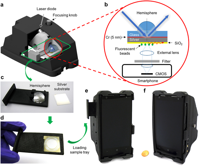
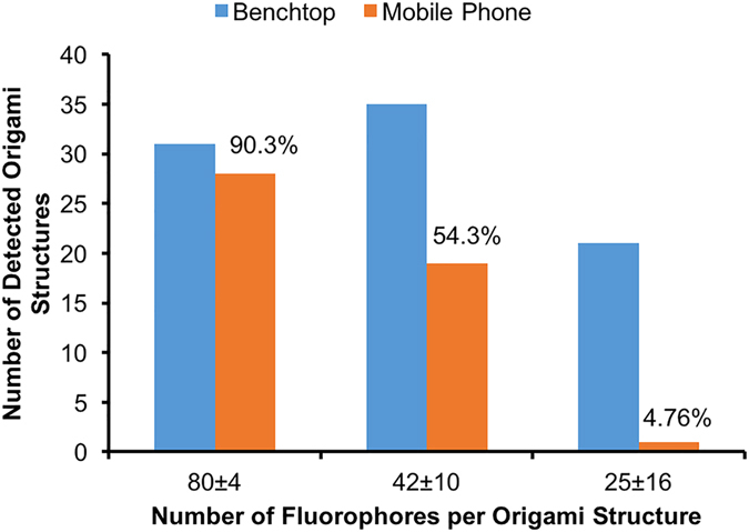
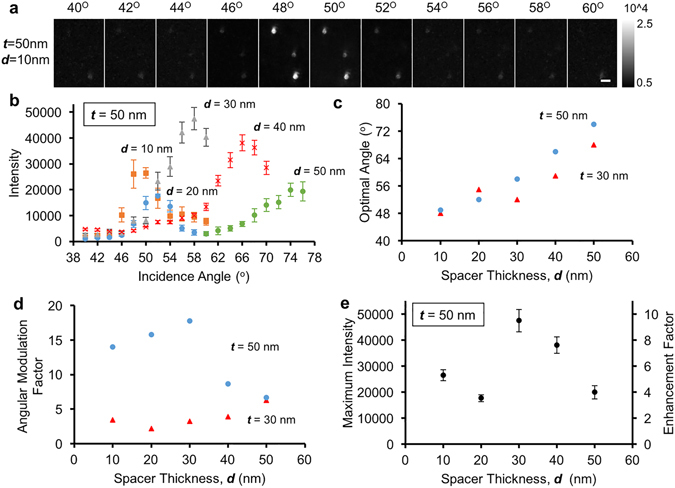
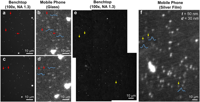
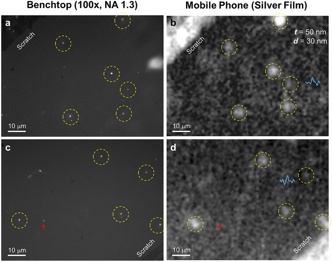
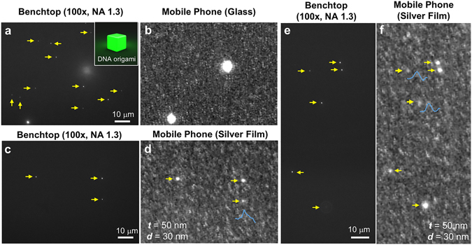
Smartphone fluorescence microscopy has various applications in point-of-care (POC) testing and diagnostics, ranging from e.g., quantification of immunoassays, detection of microorganisms, to sensing of viruses. An important need in smartphone-based microscopy and sensing techniques is to improve the detection sensitivity to enable quantification of extremely low concentrations of target molecules. Here, we demonstrate a general strategy to enhance the detection sensitivity of a smartphone-based fluorescence microscope by using surface-enhanced fluorescence (SEF) created by a thin metal-film. In this plasmonic design, the samples are placed on a silver-coated glass slide with a thin spacer, and excited by a laser-diode from the backside through a glass hemisphere, generating surface plasmon polaritons. We optimized this mobile SEF system by tuning the metal-film thickness, spacer distance, excitation angle and polarization, and achieved ~10-fold enhancement in fluorescence intensity compared to a bare glass substrate, which enabled us to image single fluorescent particles as small as 50 nm in diameter and single quantum-dots. Furthermore, we quantified the detection limit of this platform by using DNA origami-based brightness standards, demonstrating that ~80 fluorophores per diffraction-limited spot can be readily detected by our mobile microscope, which opens up new opportunities for POC diagnostics and sensing applications in resource-limited-settings.
智能手机荧光显微镜在即时检测和诊断方面有多种应用,例如免疫分析的定量、微生物的检测、病毒的传感等。基于智能手机的显微镜和传感技术的一个重要需求是提高检测灵敏度,从而能够对极低浓度的靶分子进行定量。在这里,我们展示了一种通过使用薄金属膜产生的表面增强荧光(SEF)来增强基于智能手机的荧光显微镜检测灵敏度的通用策略。在这种等离子体设计中,样品放置在带有薄间隔物的镀银玻璃载玻片上,通过玻璃半球从背面用激光二极管激发,产生表面等离子体激元。我们通过调整金属膜厚度、间隔距离、激发角度和偏振,优化了这个移动 SEF 系统,并与裸玻璃基底相比,荧光强度提高了约 10 倍,这使得我们能够对直径小至 50nm 的单个荧光颗粒和单个量子点进行成像。此外,我们使用 DNA 折纸基亮度标准来量化这个平台的检测极限,证明我们的移动显微镜可以很容易地检测到~80 个荧光团/每个衍射受限点,这为即时诊断和资源有限环境下的传感应用开辟了新的机会。