Faot F, Chatterjee M, de Camargos G V, Duyck Joke, Vandamme K
KU Leuven, University Hospitals Leuven, Department of Oral Health Sciences & Dental Clinic, BIOMAT KU Leuven & Prosthetics, Belgium.
Federal University of Pelotas, School of Dentistry, Department of Restorative Dentistry, Rio Grande do Sul, Brazil.
Bone Rep. 2015 Jan 21;2:14-24. doi: 10.1016/j.bonr.2014.10.005. eCollection 2015 Jun.
Knowledge about macro- and micro-structural characteristics may improve estimation of the quality and quantity of regenerated bone tissue. For this reason, micro-CT imaging has been applied to evaluate alveolar bone remodelling, alterations of periodontal ligament thickness and cortical and trabecular bone changes in rodent jaw bones. In this paper, we provide a systematic review on the available micro-CT literature on jaw bone micro-architecture.
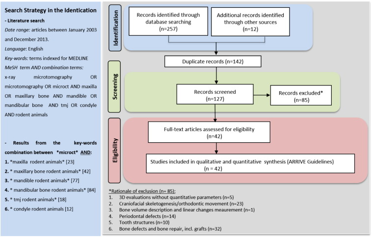
A detailed search through the PubMed database was performed. Articles published up to December 2013 and related to maxilla, mandible and condyle with quantitatively analysed bone micro-architectural parameters were considered eligible for inclusion. Two reviewers assessed the search results according to inclusion criteria designed to identify animal studies quantifying the bone micro-architecture of the jaw rodent bones in physiological or drug-induced disease status, or in response to interventions such as mechanical loading, hormonal treatment and other metabolic alterations. Finally, the reporting quality of the included publications was evaluated using the tailored ARRIVE guidelines outlined by Vignoletti and Abrahamsson (2012).
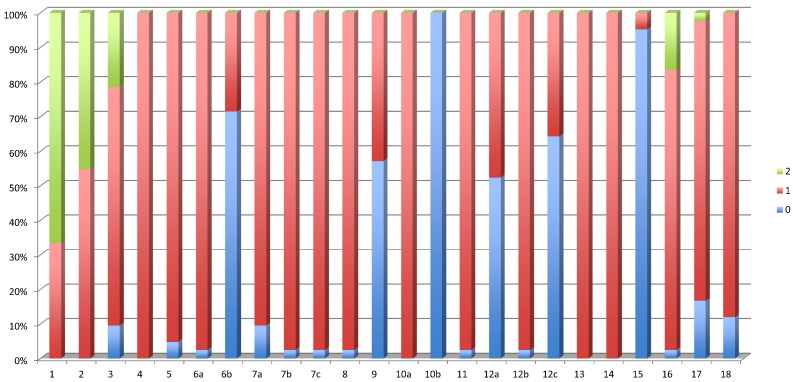
Database search, additional manual searching and assessment of the inclusion and exclusion criteria retrieved 127 potentially relevant articles. Eventually, 14 maxilla, 20 mandible and 12 condyle articles with focus on bone healing were retained, and were analysed together with 3 methodological papers. Each study was described systematically in terms of subject, experimental intervention, follow-up period, selected region of interest used in the micro-CT analysis, parameters quantified, micro-CT scanner device and software. The evidence level evaluated by the ARRIVE guidelines showed high mean scores (between 18 and 25; range: 0-25), indicating that most of the selected studies are well-reported. The major obstacles identified were related to sample size calculation, absence of adverse event descriptions, randomization or blinding procedures.
The evaluated studies are highly heterogeneous in terms of research topic and the different regions of interest. These results illustrate the need for a standardized methodology in micro-CT analysis. While the analysed studies do well according to the ARRIVE guidelines, the micro-CT procedure is often insufficiently described. Therefore we recommend to extend the ARRIVE guidelines for micro-CT studies.
了解宏观和微观结构特征可能有助于提高对再生骨组织质量和数量的评估。因此,显微计算机断层扫描(micro-CT)成像已被用于评估啮齿动物颌骨的牙槽骨重塑、牙周膜厚度变化以及皮质骨和小梁骨的改变。在本文中,我们对现有的关于颌骨微观结构的显微计算机断层扫描文献进行了系统综述。
通过PubMed数据库进行了详细检索。纳入2013年12月之前发表的、与上颌骨、下颌骨和髁突相关且对骨微观结构参数进行了定量分析的文章。两名评审员根据纳入标准评估检索结果,这些标准旨在识别量化生理或药物诱导疾病状态下或对机械负荷、激素治疗和其他代谢改变等干预措施作出反应时啮齿动物颌骨骨微观结构的动物研究。最后,使用Vignoletti和Abrahamsson(2012年)概述的定制ARRIVE指南评估纳入出版物的报告质量。
数据库检索、额外的手动检索以及纳入和排除标准评估共检索到127篇潜在相关文章。最终,保留了14篇关注上颌骨、20篇关注下颌骨和12篇关注髁突且聚焦于骨愈合的文章,并与3篇方法学论文一起进行分析。每项研究都从研究对象、实验干预、随访期、显微计算机断层扫描分析中使用的选定感兴趣区域、量化参数、显微计算机断层扫描仪设备和软件等方面进行了系统描述。ARRIVE指南评估的证据水平显示平均得分较高(介于18至25分之间;范围:0 - 25分),表明大多数选定研究报告良好。确定的主要障碍与样本量计算、不良事件描述缺失、随机化或盲法程序有关。
所评估的研究在研究主题和不同感兴趣区域方面高度异质。这些结果表明在显微计算机断层扫描分析中需要标准化方法。虽然根据ARRIVE指南分析的研究表现良好,但显微计算机断层扫描程序的描述往往不够充分。因此,我们建议扩展针对显微计算机断层扫描研究的ARRIVE指南。