IEEE J Biomed Health Inform. 2017 Nov;21(6):1607-1616. doi: 10.1109/JBHI.2017.2704614. Epub 2017 May 16.
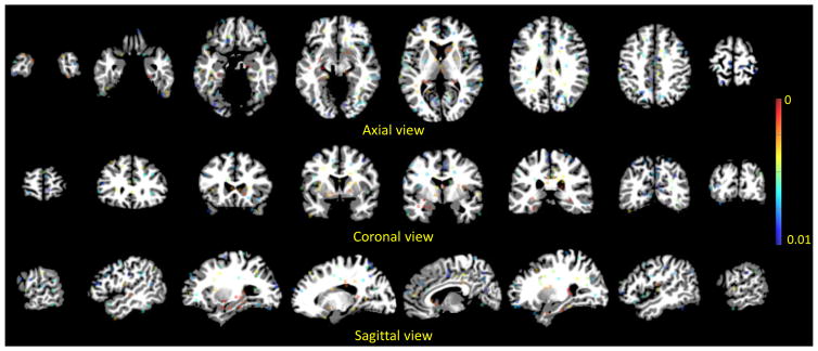
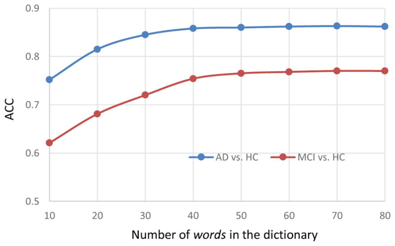
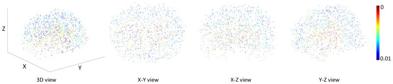
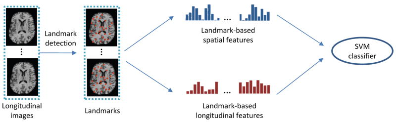
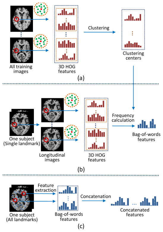
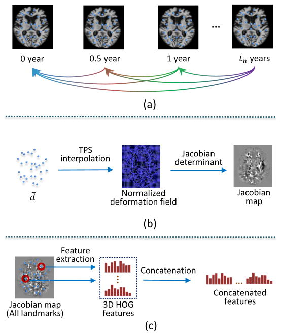
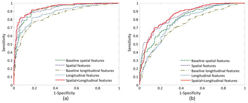
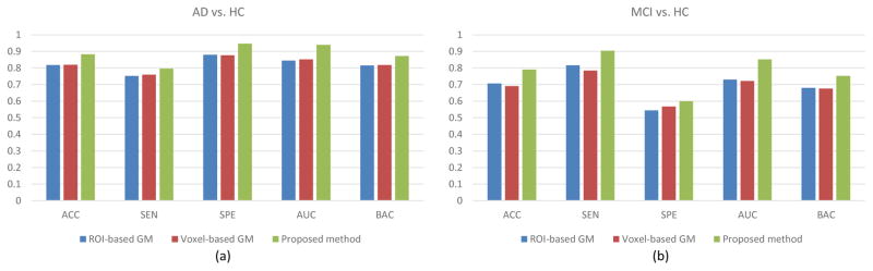
Structural magnetic resonance imaging (MRI) has been proven to be an effective tool for Alzheimer's disease (AD) diagnosis. While conventional MRI-based AD diagnosis typically uses images acquired at a single time point, a longitudinal study is more sensitive in detecting early pathological changes of AD, making it more favorable for accurate diagnosis. In general, there are two challenges faced in MRI-based diagnosis. First, extracting features from structural MR images requires time-consuming nonlinear registration and tissue segmentation, whereas the longitudinal study with involvement of more scans further exacerbates the computational costs. Moreover, the inconsistent longitudinal scans (i.e., different scanning time points and also the total number of scans) hinder extraction of unified feature representations in longitudinal studies. In this paper, we propose a landmark-based feature extraction method for AD diagnosis using longitudinal structural MR images, which does not require nonlinear registration or tissue segmentation in the application stage and is also robust to inconsistencies among longitudinal scans. Specifically, first, the discriminative landmarks are automatically discovered from the whole brain using training images, and then efficiently localized using a fast landmark detection method for testing images, without the involvement of any nonlinear registration and tissue segmentation; and second, high-level statistical spatial features and contextual longitudinal features are further extracted based on those detected landmarks, which can characterize spatial structural abnormalities and longitudinal landmark variations. Using these spatial and longitudinal features, a linear support vector machine is finally adopted to distinguish AD subjects or mild cognitive impairment (MCI) subjects from healthy controls (HCs). Experimental results on the Alzheimer's Disease Neuroimaging Initiative database demonstrate the superior performance and efficiency of the proposed method, with classification accuracies of 88.30% for AD versus HC and 79.02% for MCI versus HC, respectively.
结构磁共振成像(MRI)已被证明是诊断阿尔茨海默病(AD)的有效工具。虽然基于常规 MRI 的 AD 诊断通常使用单个时间点采集的图像,但纵向研究在检测 AD 的早期病理变化方面更为敏感,因此更有利于准确诊断。一般来说,基于 MRI 的诊断面临两个挑战。首先,从结构 MRI 图像中提取特征需要耗时的非线性配准和组织分割,而涉及更多扫描的纵向研究进一步增加了计算成本。此外,不一致的纵向扫描(即不同的扫描时间点和扫描总数)阻碍了在纵向研究中提取统一的特征表示。在本文中,我们提出了一种基于标志点的特征提取方法,用于使用纵向结构 MRI 进行 AD 诊断,该方法在应用阶段不需要非线性配准或组织分割,并且对纵向扫描的不一致性具有鲁棒性。具体来说,首先,使用训练图像从整个大脑中自动发现有区分力的标志点,然后使用快速标志点检测方法在测试图像中有效地定位标志点,而无需任何非线性配准和组织分割;其次,基于那些检测到的标志点进一步提取高级统计空间特征和上下文纵向特征,这些特征可以表征空间结构异常和纵向标志点变化。最后,使用这些空间和纵向特征,采用线性支持向量机来区分 AD 患者或轻度认知障碍(MCI)患者与健康对照组(HC)。在阿尔茨海默病神经影像学倡议数据库上的实验结果证明了所提出方法的优越性能和效率,AD 与 HC 之间的分类准确率为 88.30%,MCI 与 HC 之间的分类准确率为 79.02%。