Ihle-Hansen Hege, Hagberg Guri, Fure Brynjar, Thommessen Bente, Fagerland Morten W, Øksengård Anne R, Engedal Knut, Selnes Per
Department of Internal medicine, Vestre Viken Hospital Trust, Baerum Hospital, Norway, 3004, Drammen, Norway.
Norwegian Knowledge Centre for the Health Services, Oslo, Norway.
BMC Neurol. 2017 Jun 5;17(1):107. doi: 10.1186/s12883-017-0890-6.
Although the most serious consequence of neuronal ischemia is acute neuronal death, mounting evidence suggests similarities between stroke and neurodegenerative disease. Brain atrophy visualized on structural MRI and pathological cerebrospinal fluid (CSF) concentrations of microtubule-associated protein tau (T-tau) and phosphorylated microtubule-associated protein tau indicate neurofibrillary degeneration. We aimed to explore the association between CSF T-tau and brain atrophy 1 year post-stroke.
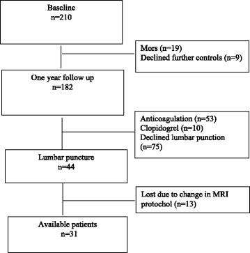
We included 210 patients with first-ever ischemic stroke or transitory ischemic attack without pre-existing cognitive impairment. After 12 months, subjects underwent MRI, and CSF biomarkers were assessed. Using SIENAX (part of FSL), ventricular CSF volume and total brain volume were estimated and normalized for subject head size. The association between T-tau as explanatory variable and ventricular and total brain volume as outcome variables were studied using linear regression.
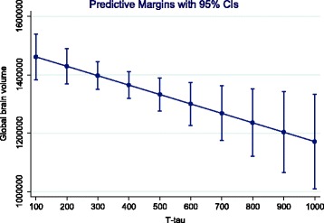
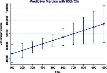
One hundred eighty-two patients completed the follow-up. Forty-four had a lumbar puncture. Of these, 31 had their MRI with identical scan parameters. Mean age was 70.2 years (SD 11.7). Ventricular volume on MRI was significantly associated with age, but not with gender. In the multiple regression model, there was a significant association between T-tau and both ventricular (beta 0.44, 95% CI 376.3, 394.9, p = 0.021) and global brain volume (beta -0.50, 95% CI -565.9, -78.3, p = 0.011). There was no significant association between CSF T-tau 1 year post-stroke and baseline volumes.
T-tau measured 1 year post-stroke is associated with measures of brain atrophy. The findings indicate that acute stroke may enhance or trigger tau-linked neurodegeneration with loss of neurons.
Clinicaltrials.gov NCT00506818 , July 23, 2007. Inclusion from February 2007, randomization and intervention from May 2007 and trial registration in July 2007.
尽管神经元缺血最严重的后果是急性神经元死亡,但越来越多的证据表明中风与神经退行性疾病之间存在相似之处。结构磁共振成像(MRI)显示的脑萎缩以及病理性脑脊液(CSF)中微管相关蛋白tau(T-tau)和磷酸化微管相关蛋白tau的浓度表明存在神经原纤维变性。我们旨在探讨中风后1年脑脊液T-tau与脑萎缩之间的关联。
我们纳入了210例首次发生缺血性中风或短暂性脑缺血发作且无既往认知障碍的患者。12个月后,受试者接受了MRI检查,并对脑脊液生物标志物进行了评估。使用SIENAX(FSL的一部分)估算脑室脑脊液体积和全脑体积,并根据受试者头部大小进行标准化。以T-tau作为解释变量,以脑室和全脑体积作为结果变量,采用线性回归研究它们之间的关联。
182例患者完成了随访。44例进行了腰椎穿刺。其中,31例接受了参数相同的MRI检查。平均年龄为70.2岁(标准差11.7)。MRI上的脑室体积与年龄显著相关,但与性别无关。在多元回归模型中,T-tau与脑室(β0.44,95%可信区间376.3,394.9,p = 0.021)和全脑体积(β -0.50,95%可信区间-565.9,-78.3,p = 0.011)均存在显著关联。中风后1年脑脊液T-tau与基线体积之间无显著关联。
中风后1年测得的T-tau与脑萎缩指标相关。研究结果表明,急性中风可能会增强或引发与tau相关的神经退行性变并导致神经元丢失。
Clinicaltrials.gov NCT00506818,2007年7月23日。2007年2月开始纳入,2007年5月进行随机分组和干预,2007年7月进行试验注册。