Moradi Khaniabadi Pegah, Shahbazi-Gahrouei Daryoush, Malik Shah Abdul Majid Amin, Suhaimi Jaafar Mohammad, Moradi Khaniabadi Bita, Shahbazi-Gahrouei Saghar
School of Physics, Universiti Sains Malaysia11800, Pulau Penang, Malaysia.
Department of Medical Physics, School of Medicine, Isfahan University of Medical Sciences, Isfahan, Iran.
Iran Biomed J. 2017 Nov;21(6):360-8. doi: 10.18869/acadpub.ibj.21.6.360. Epub 2017 Jun 11.
Magnetic resonance imaging (MRI) plays an essential role in molecular imaging by delivering the contrast agent into targeted cancer cells. The aim of this study was to evaluate the C595 monoclonal antibody-conjugated superparamagnetic iron oxide nanoparticles (SPIONs-C595) for the detection of breast cancer cell (MCF-7).
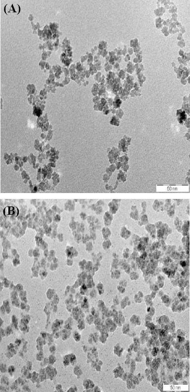
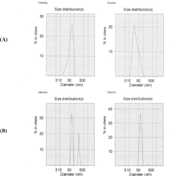
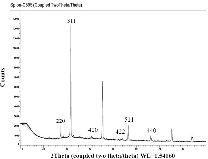
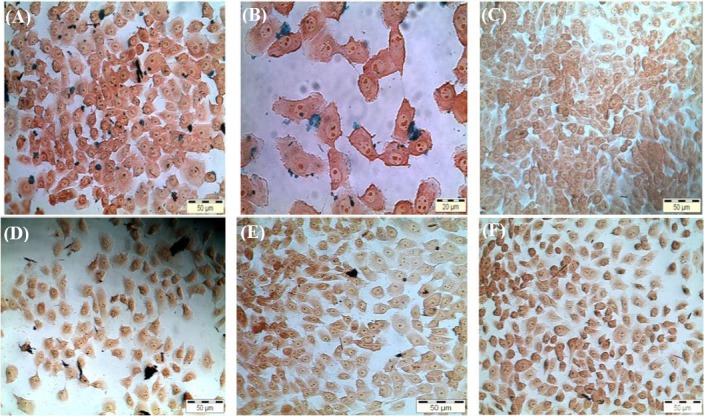
The conjugation of monoclonal antibody and nanoparticles was confirmed using X-ray diffraction, transmission electron microscopy, and photon correlation spectroscopy. The selectivity of the nanoprobe for breast cancer cells (MCF-7) was obtained by Prussian blue, atomic emission spectroscopy, and MRI relaxometry.
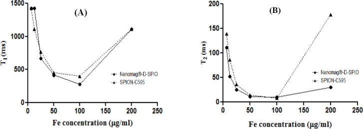
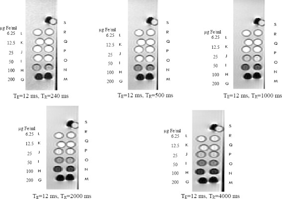
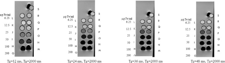
The in vitro MRI showed that T2 relaxation time will be reduced 76% when using T2-weighed magnetic resonance images compared to the control group (untreated cells) at the dose of 200 μg Fe/ml, as the optimum dose. In addition, the results showed the high uptake of nanoprobe into MCF-7 cancer cells.
The SPIONs-C595 nanoprobe has potential for the detection of specific breast cancer.
磁共振成像(MRI)通过将造影剂输送到靶向癌细胞中,在分子成像中发挥着重要作用。本研究的目的是评估C595单克隆抗体偶联的超顺磁性氧化铁纳米颗粒(SPIONs-C595)用于检测乳腺癌细胞(MCF-7)。
使用X射线衍射、透射电子显微镜和光子相关光谱法确认单克隆抗体与纳米颗粒的偶联。通过普鲁士蓝、原子发射光谱法和MRI弛豫测量法获得纳米探针对乳腺癌细胞(MCF-7)的选择性。
体外MRI显示,在200μg Fe/ml的剂量下,作为最佳剂量,与对照组(未处理细胞)相比,使用T2加权磁共振图像时,T2弛豫时间将减少76%。此外,结果显示纳米探针对MCF-7癌细胞的摄取量很高。
SPIONs-C595纳米探针具有检测特定乳腺癌的潜力。