Moradi Khaniabadi Pegah, Shahbazi-Gahrouei Daryoush, Jaafar Mohammad Suhaimi, Majid Amin Malik Shah Abdul, Moradi Khaniabadi Bita, Shahbazi-Gahrouei Saghar
Faculty of Physics, University Sains Malaysia, Pulau Penang, Malaysia.
Department of Medical Physics, Faculty of Medicine, Isfahan University of Medical Sciences, Isfahan, Iran.
Avicenna J Med Biotechnol. 2017 Oct-Dec;9(4):181-188.
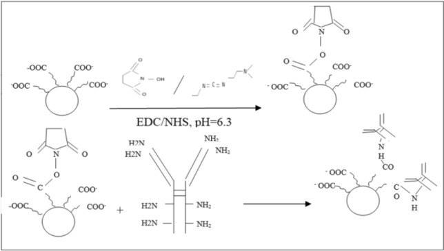
Advances of nanotechnology have led to the development of nano-materials with both potential diagnostic and therapeutic applications. Among them, Super Paramagnetic Iron Oxide Nanoparticles (SPIONs) have received particular attention. Modified EDC coupling fraction was used to fabricate the SPION-C595 as an MR imaging contrast agent for breast cancer detection in early stages.
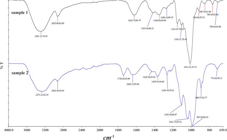
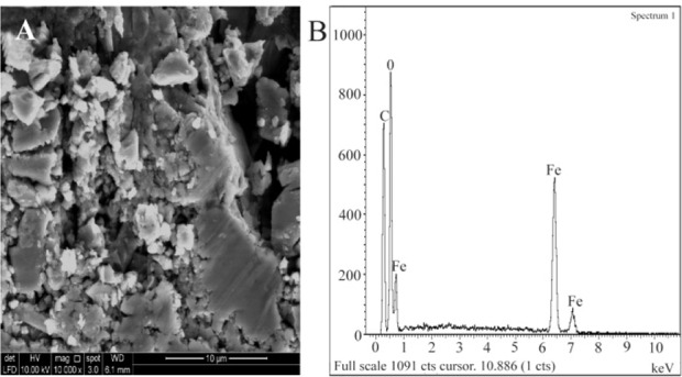
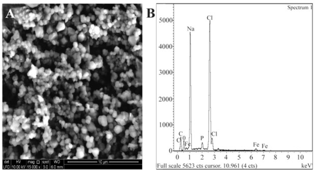
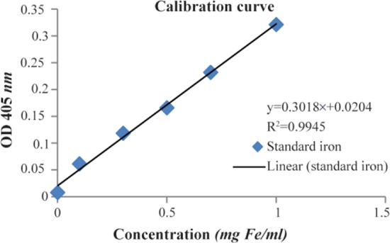
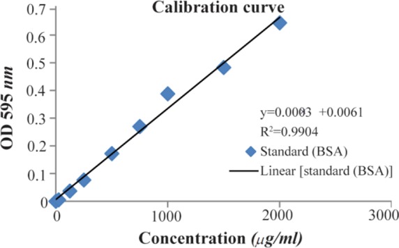
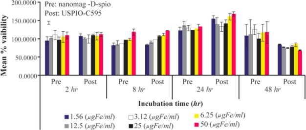
Nanoprobe characterization was confirmed using Fourier Transform Infrared Spectroscopy (FT-IR), Scanning Electron Microscopy with Energy Dispersive X-Ray Spectroscopy (SEM-EDAX), and Photon Correlation Spectroscopy (PCS). Protein and iron concentration of nanoprobe was examined by standard method. MTT assay was performed to evaluate the cytotoxicity of the nanoprobe in breast cancer cell line (MCF-7). T-weighted MR imaging was performed to evaluate the signal enhancement on T relaxation time of nanoprobe using spin-echo pulse sequence.
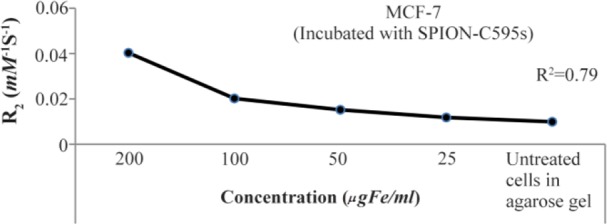
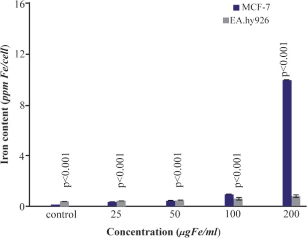
As results showed, SPIONs-C595 provided active targeting of breast cancer cell (MCF-7) at a final concentration of 600 . The final concentration of protein was calculated to be at 0.78 . The hydrodynamic size of the nanoprobe was 87.4±0.7 . The MR imaging results showed a good reduction of T relaxation rates for the highest dose of SPIONs-C595.
Based on the results, SPIONs-C595 nanoprobe has a potential in T-weighted MR imaging contrast agent for breast cancer cell (MCF-7) detection.
纳米技术的进步促使了具有潜在诊断和治疗应用的纳米材料的发展。其中,超顺磁性氧化铁纳米颗粒(SPIONs)受到了特别关注。采用改良的EDC偶联分数制备了SPION-C595,作为用于早期乳腺癌检测的磁共振成像造影剂。
使用傅里叶变换红外光谱(FT-IR)、带能谱仪的扫描电子显微镜(SEM-EDAX)和光子相关光谱(PCS)对纳米探针进行表征。通过标准方法检测纳米探针的蛋白质和铁浓度。进行MTT试验以评估纳米探针对乳腺癌细胞系(MCF-7)的细胞毒性。使用自旋回波脉冲序列进行T加权磁共振成像,以评估纳米探针在T弛豫时间上的信号增强。
结果显示,在最终浓度为600时,SPIONs-C595对乳腺癌细胞(MCF-7)具有主动靶向作用。计算得出蛋白质的最终浓度为0.78。纳米探针的流体动力学尺寸为87.4±0.7。磁共振成像结果表明,最高剂量的SPIONs-C595能使T弛豫率显著降低。
基于这些结果,SPIONs-C595纳米探针在用于检测乳腺癌细胞(MCF-7)的T加权磁共振成像造影剂方面具有潜力。