• 文献检索
  • 文档翻译
  • 深度研究
  • 学术资讯
  • Suppr Zotero 插件Zotero 插件
  • 邀请有礼
  • 套餐&价格
  • 历史记录
应用&插件
Suppr Zotero 插件Zotero 插件浏览器插件Mac 客户端Windows 客户端微信小程序
定价
高级版会员购买积分包购买API积分包
服务
文献检索文档翻译深度研究API 文档MCP 服务
关于我们
关于 Suppr公司介绍联系我们用户协议隐私条款
关注我们

Suppr 超能文献

核心技术专利:CN118964589B侵权必究
粤ICP备2023148730 号-1Suppr @ 2026

文献检索

告别复杂PubMed语法,用中文像聊天一样搜索,搜遍4000万医学文献。AI智能推荐,让科研检索更轻松。

立即免费搜索

文件翻译

保留排版,准确专业,支持PDF/Word/PPT等文件格式,支持 12+语言互译。

免费翻译文档

深度研究

AI帮你快速写综述,25分钟生成高质量综述,智能提取关键信息,辅助科研写作。

立即免费体验

问答:扩展显微镜技术

Q&A: Expansion microscopy.

作者信息

Gao Ruixuan, Asano Shoh M, Boyden Edward S

机构信息

Media Lab, Massachusetts Institute of Technology (MIT), Cambridge, MA, USA.

Internal Medicine Research Unit, Pfizer Inc., Cambridge, MA, USA.

出版信息

BMC Biol. 2017 Jun 19;15(1):50. doi: 10.1186/s12915-017-0393-3.

DOI:10.1186/s12915-017-0393-3
PMID:28629474
原文链接:https://pmc.ncbi.nlm.nih.gov/articles/PMC5474863/
Abstract

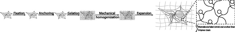
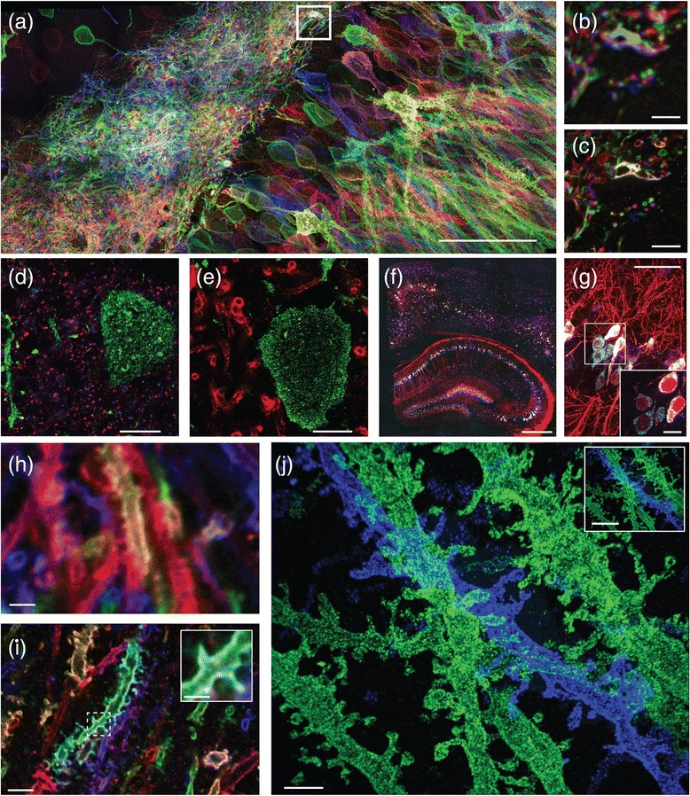
Expansion microscopy (ExM) is a recently invented technology that uses swellable charged polymers, synthesized densely and with appropriate topology throughout a preserved biological specimen, to physically magnify the specimen 100-fold in volume, or more, in an isotropic fashion. ExM enables nanoscale resolution imaging of preserved samples on inexpensive, fast, conventional microscopes. How does ExM work? How good is its performance? How do you get going on using it? In this Q&A, we provide the answers to these and other questions about this new and rapidly spreading toolbox.

摘要

扩展显微镜技术(ExM)是一项最近发明的技术,它使用可膨胀的带电聚合物,在整个保存的生物样本中密集合成并具有适当的拓扑结构,以各向同性的方式将样本的体积物理放大100倍或更多。ExM能够在廉价、快速的传统显微镜上对保存的样本进行纳米级分辨率成像。ExM是如何工作的?它的性能如何?如何开始使用它?在这个问答中,我们提供了关于这个新的且迅速传播的工具包的这些问题及其他问题的答案。

https://cdn.ncbi.nlm.nih.gov/pmc/blobs/c857/5474863/7c4b9c4e852d/12915_2017_393_Fig3_HTML.jpg
https://cdn.ncbi.nlm.nih.gov/pmc/blobs/c857/5474863/cdd61b3c7401/12915_2017_393_Fig1_HTML.jpg
https://cdn.ncbi.nlm.nih.gov/pmc/blobs/c857/5474863/2db8f773f385/12915_2017_393_Fig2_HTML.jpg
https://cdn.ncbi.nlm.nih.gov/pmc/blobs/c857/5474863/7c4b9c4e852d/12915_2017_393_Fig3_HTML.jpg
https://cdn.ncbi.nlm.nih.gov/pmc/blobs/c857/5474863/cdd61b3c7401/12915_2017_393_Fig1_HTML.jpg
https://cdn.ncbi.nlm.nih.gov/pmc/blobs/c857/5474863/2db8f773f385/12915_2017_393_Fig2_HTML.jpg
https://cdn.ncbi.nlm.nih.gov/pmc/blobs/c857/5474863/7c4b9c4e852d/12915_2017_393_Fig3_HTML.jpg

相似文献

1
Q&A: Expansion microscopy.问答:扩展显微镜技术
BMC Biol. 2017 Jun 19;15(1):50. doi: 10.1186/s12915-017-0393-3.
2
Expansion Microscopy for Beginners: Visualizing Microtubules in Expanded Cultured HeLa Cells.初学者的扩展显微镜技术:在扩展培养的 HeLa 细胞中可视化微管。
Curr Protoc Neurosci. 2020 Jun;92(1):e96. doi: 10.1002/cpns.96.
3
Iterative expansion microscopy.迭代扩展显微镜技术。
Nat Methods. 2017 Jun;14(6):593-599. doi: 10.1038/nmeth.4261. Epub 2017 Apr 17.
4
Expansion Microscopy: Protocols for Imaging Proteins and RNA in Cells and Tissues.扩展显微镜技术:细胞和组织中蛋白质与RNA成像方案
Curr Protoc Cell Biol. 2018 Sep;80(1):e56. doi: 10.1002/cpcb.56. Epub 2018 Aug 2.
5
Basics of Expansion Microscopy.扩展显微镜基础
Curr Protoc Cytom. 2019 Dec;91(1):e67. doi: 10.1002/cpcy.67.
6
Expansion microscopy: principles and uses in biological research.扩展显微镜:在生物研究中的原理和应用。
Nat Methods. 2019 Jan;16(1):33-41. doi: 10.1038/s41592-018-0219-4. Epub 2018 Dec 20.
7
Optical imaging. Expansion microscopy.光学成像。扩展显微镜技术。
Science. 2015 Jan 30;347(6221):543-8. doi: 10.1126/science.1260088. Epub 2015 Jan 15.
8
Plasmon-Enhanced Expansion Microscopy.等离子体增强扩展显微镜。
Nano Lett. 2023 Jun 28;23(12):5654-5662. doi: 10.1021/acs.nanolett.3c01256. Epub 2023 Jun 12.
9
Expansion Microscopy with Multifunctional Polymer Dots.多功能聚合物点的扩展显微镜技术。
Adv Mater. 2021 Jun;33(25):e2007854. doi: 10.1002/adma.202007854. Epub 2021 May 14.
10
Nanoscale imaging using differential expansion microscopy.使用差分扩展显微镜进行纳米级成像。
Histochem Cell Biol. 2020 Jun;153(6):469-480. doi: 10.1007/s00418-020-01869-7. Epub 2020 Mar 19.

引用本文的文献

1
Expansion and fluctuations-enhanced microscopy for nanoscale molecular profiling of cells and tissues.用于细胞和组织纳米级分子分析的扩展与波动增强显微镜技术。
Nat Protoc. 2025 Jul 2. doi: 10.1038/s41596-025-01178-0.
2
Rapid lightsheet fluorescence imaging of whole Drosophila brains at nanoscale resolution by potassium acrylate-based expansion microscopy.通过基于丙烯酸钾的膨胀显微镜对果蝇全脑进行纳米级分辨率的快速光片荧光成像。
Nat Commun. 2024 Dec 30;15(1):10911. doi: 10.1038/s41467-024-55305-8.
3
Refining Biologically Inconsistent Segmentation Masks with Masked Autoencoders.

本文引用的文献

1
Iterative expansion microscopy.迭代扩展显微镜技术。
Nat Methods. 2017 Jun;14(6):593-599. doi: 10.1038/nmeth.4261. Epub 2017 Apr 17.
2
The tetrameric kinesin Kif25 suppresses pre-mitotic centrosome separation to establish proper spindle orientation.四聚体驱动蛋白Kif25抑制有丝分裂前中心体分离以建立正确的纺锤体方向。
Nat Cell Biol. 2017 Apr;19(4):384-390. doi: 10.1038/ncb3486. Epub 2017 Mar 6.
3
Influenza virus exploits tunneling nanotubes for cell-to-cell spread.流感病毒利用隧道纳米管进行细胞间传播。
使用掩码自动编码器优化生物学上不一致的分割掩码。
Conf Comput Vis Pattern Recognit Workshops. 2024 Jun 17:6904-6912. doi: 10.1109/CVPRW63382.2024.00684.
4
3D-Aligner: advanced computational tool for correcting image distortion in expansion microscopy.3D-Aligner:用于校正扩展显微镜图像失真的高级计算工具。
Commun Biol. 2024 Oct 15;7(1):1325. doi: 10.1038/s42003-024-07035-2.
5
Expansion Microscopy of Synaptic Contacts on the Mauthner Cells of Larval Zebrafish.斑马鱼幼体Mauthner细胞突触接触的扩展显微镜观察
Bio Protoc. 2024 Sep 20;14(18):e5067. doi: 10.21769/BioProtoc.5067.
6
3D-Aligner: An advanced computational tool designed to correct image distortion in expansion microscopy for precise 3D reconstitution and quantitative analysis.3D对齐器:一种先进的计算工具,旨在校正扩展显微镜中的图像失真,以实现精确的三维重建和定量分析。
bioRxiv. 2024 Aug 30:2024.08.28.610199. doi: 10.1101/2024.08.28.610199.
7
Nanoscale insights into hematology: super-resolved imaging on blood cell structure, function, and pathology.纳米尺度洞察血液学:血细胞结构、功能和病理学的超高分辨率成像。
J Nanobiotechnology. 2024 Jun 24;22(1):363. doi: 10.1186/s12951-024-02605-2.
8
Pre-gelation staining expansion microscopy for visualisation of the Plasmodium liver stage.胶前染色扩展显微镜用于可视化疟原虫肝期。
J Cell Sci. 2023 Nov 15;136(22). doi: 10.1242/jcs.261377. Epub 2023 Nov 30.
9
Expansion microscopy in honeybee brains for high-resolution neuroanatomical analyses in social insects.在蜜蜂大脑中进行扩展显微镜技术,以进行社会昆虫的高分辨率神经解剖学分析。
Cell Tissue Res. 2023 Sep;393(3):489-506. doi: 10.1007/s00441-023-03803-4. Epub 2023 Jul 8.
10
Therapeutic Ultrasound for Topical Corneal Delivery of Macromolecules.治疗超声促进大分子眼表给药
Transl Vis Sci Technol. 2022 Aug 1;11(8):23. doi: 10.1167/tvst.11.8.23.
Sci Rep. 2017 Jan 6;7:40360. doi: 10.1038/srep40360.
4
High-performance multiplexed fluorescence in situ hybridization in culture and tissue with matrix imprinting and clearing.利用基质印记和透明化技术在培养物和组织中进行的高性能多重荧光原位杂交。
Proc Natl Acad Sci U S A. 2016 Dec 13;113(50):14456-14461. doi: 10.1073/pnas.1617699113. Epub 2016 Nov 22.
5
In Situ Transcription Profiling of Single Cells Reveals Spatial Organization of Cells in the Mouse Hippocampus.单细胞原位转录谱分析揭示小鼠海马体中细胞的空间组织
Neuron. 2016 Oct 19;92(2):342-357. doi: 10.1016/j.neuron.2016.10.001.
6
Striosome-dendron bouquets highlight a unique striatonigral circuit targeting dopamine-containing neurons.纹状体小体-树突束突出了一个针对含多巴胺神经元的独特纹状体黑质回路。
Proc Natl Acad Sci U S A. 2016 Oct 4;113(40):11318-11323. doi: 10.1073/pnas.1613337113. Epub 2016 Sep 19.
7
smiFISH and FISH-quant - a flexible single RNA detection approach with super-resolution capability.smiFISH和FISH定量——一种具有超分辨率能力的灵活单RNA检测方法。
Nucleic Acids Res. 2016 Dec 15;44(22):e165. doi: 10.1093/nar/gkw784. Epub 2016 Sep 5.
8
Multiplexed and scalable super-resolution imaging of three-dimensional protein localization in size-adjustable tissues.在尺寸可调组织中对三维蛋白质定位进行多重且可扩展的超分辨率成像。
Nat Biotechnol. 2016 Sep;34(9):973-81. doi: 10.1038/nbt.3641. Epub 2016 Jul 25.
9
Nanoscale imaging of RNA with expansion microscopy.利用扩张显微镜对RNA进行纳米级成像。
Nat Methods. 2016 Aug;13(8):679-84. doi: 10.1038/nmeth.3899. Epub 2016 Jul 4.
10
Protein-retention expansion microscopy of cells and tissues labeled using standard fluorescent proteins and antibodies.使用标准荧光蛋白和抗体标记的细胞和组织的蛋白质保留扩展显微镜技术。
Nat Biotechnol. 2016 Sep;34(9):987-92. doi: 10.1038/nbt.3625. Epub 2016 Jul 4.