Gong Zongrong, Miao Ruixue, Shu Min, Zhu Yu, Wen Yang, Guo Qin, Liao Qiong, Wan Chaomin
Department of Pediatric Infectious Disease, West China Second University Hospital/West China Women's and Children's Hospital, Sichuan University, Chengdu, Key Laboratory of Birth Defects and Related Diseases of Women and Children, Ministry of Education (Sichuan University) China.
Medicine (Baltimore). 2017 Jun;96(25):e7265. doi: 10.1097/MD.0000000000007265.
Paragonimiasis infection has no specific symptoms or typical radiologic findings, leading to the possibility of misdiagnosis. Thus, the objective of this study was to analyze clinical and radiological features, and treatment outcome of paragonimiasis in children in Southwest China to improve the awareness of this disease.
We retrospectively reviewed the records of children diagnosed with paragonimiasis in West China Second University Hospital between 2005 and 2016. The confirmed diagnosis of paragonimiasis was based on epidemiology history and seropositivity for paragonimiasis and/or detection of paragonimus eggs. Clinical, laboratory, and imaging findings of patients were examined in order to summarize risk factors, clinical characteristics, and treatment outcomes of these patients.
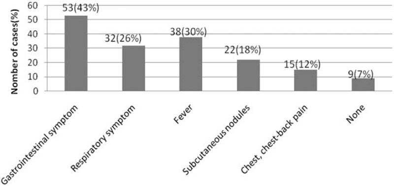
A total of 123 patients were included; of them 112 (91.1%) lived in villages and 72 (58.5%) had a history of consuming freshwater crabs. Patients with paragonimiasis most frequently showed respiratory symptoms, including cough (26.0%, 32/123) and tachypnea (16.3%, 20/123), and gastrointestinal symptoms, including abdominal pain (26.8%, 33/123), abdominal distention (22.8%, 28/123), and vomiting (13.0%, 16/123). Laboratory examination showed elevated white blood cell (WBC) counts in the peripheral blood in 89 (72.4%) patients and eosinophilia in 102 (82.9%) patients. Tuberculosis (TB) coinfection was found in 4 (3.3%) patients. Main imaging findings included: effusions (90.4%), lymphadenopathy (40.4%), pulmonary ground-glass opacities (36.2%), cystic lesions (18.1%), and pleural thickening (17.0%). Twenty-nine patients (23.6%) received more than 1 course of praziquantel (PZQ). Additionally, 4 (19.0%) of 21 patients who were discharged from the hospital without complete treatment required rehospitalization for residual serous effusions. Moreover, patients from pericardial effusion group showed longer hospital stays and less elevated WBC counts than those from nonpericardial effusion group.
Paragonimiasis should be considered in patients from endemic areas, especially in those with gastrointestinal and/or respiratory symptoms, elevated WBC count, eosinophilia, and serous effusions. Additionally, longer hospital stay may be necessary in cases of paragonimiasis associated with pericardial effusions.
肺吸虫病感染无特异性症状或典型的影像学表现,导致误诊的可能性。因此,本研究的目的是分析中国西南部儿童肺吸虫病的临床和放射学特征及治疗结果,以提高对该病的认识。
我们回顾性分析了2005年至2016年在西部战区总医院被诊断为肺吸虫病的儿童病历。肺吸虫病的确诊基于流行病学史、肺吸虫病血清学阳性和/或肺吸虫虫卵检测。对患者的临床、实验室和影像学检查结果进行分析,以总结这些患者的危险因素、临床特征和治疗结果。
共纳入123例患者;其中112例(91.1%)居住在农村,72例(58.5%)有食用淡水蟹史。肺吸虫病患者最常见的症状为呼吸道症状,包括咳嗽(26.0%,32/123)和呼吸急促(16.3%,20/123),以及胃肠道症状,包括腹痛(26.8%,33/123)、腹胀(22.8%,28/123)和呕吐(13.0%,16/123)。实验室检查显示,89例(72.4%)患者外周血白细胞(WBC)计数升高,102例(82.9%)患者嗜酸性粒细胞增多。4例(3.3%)患者合并肺结核(TB)感染。主要影像学表现包括:胸腔积液(90.4%)、淋巴结肿大(40.4%)、肺部磨玻璃影(36.2%)、囊性病变(18.1%)和胸膜增厚(17.0%)。29例(23.6%)患者接受了超过1个疗程的吡喹酮(PZQ)治疗。此外,21例未接受完整治疗出院的患者中有4例(19.0%)因残留胸腔积液需要再次住院。此外,心包积液组患者的住院时间比非心包积液组患者长,WBC计数升高幅度较小。
来自流行地区的患者,尤其是有胃肠道和/或呼吸道症状、WBC计数升高、嗜酸性粒细胞增多和胸腔积液的患者,应考虑肺吸虫病。此外,与心包积液相关的肺吸虫病患者可能需要更长的住院时间。