Kaese Sven, Larbig Robert, Rohrbeck Matthias, Frommeyer Gerrit, Dechering Dirk, Olligs Jan, Schönhofer-Merl Sabine, Wessely Rainer, Klingel Karin, Seebohm Guiscard, Eckardt Lars
Division of Electrophysiology, Department of Cardiovascular Medicine, University of Münster, Münster, Germany.
The IfGH-Myocellular Electrophysiology, Department of Cardiovascular Medicine, University of Münster, Münster, Germany.
PLoS One. 2017 Jun 23;12(6):e0180029. doi: 10.1371/journal.pone.0180029. eCollection 2017.
Coxsackievirus B3 (CVB3) is known to induce acute and chronic myocarditis. Most infections are clinically unapparent but some patients suffer from ventricular arrhythmias (VA) and sudden cardiac death (SCD). Studies showed that acute CVB3 infection may cause impaired function of cardiac ion channels, creating a proarrhythmic substrate. However, it is unknown whether low level CVB3+ expression in myocytes may cause altered cardiac electrophysiology leading to VA.
Cellular electrophysiology was used to analyze cellular action potentials (APs) and occurrence of afterdepolarizations from isolated cardiomyocytes of wildtype (WT) and transgenic CVB3ΔVP0 (CVB3+) mice. Further, we studied surface ECGs, monophasic APs, ventricular effective refractory period (VERP) and inducibility of VAs in Langendorff-perfused whole hearts. All used cardiomyocytes and whole hearts originated from male mice.
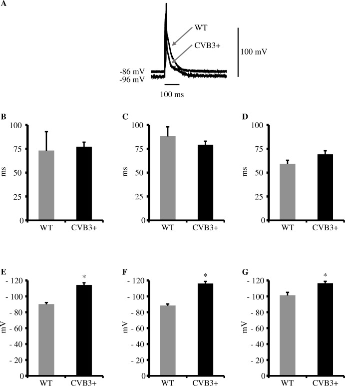
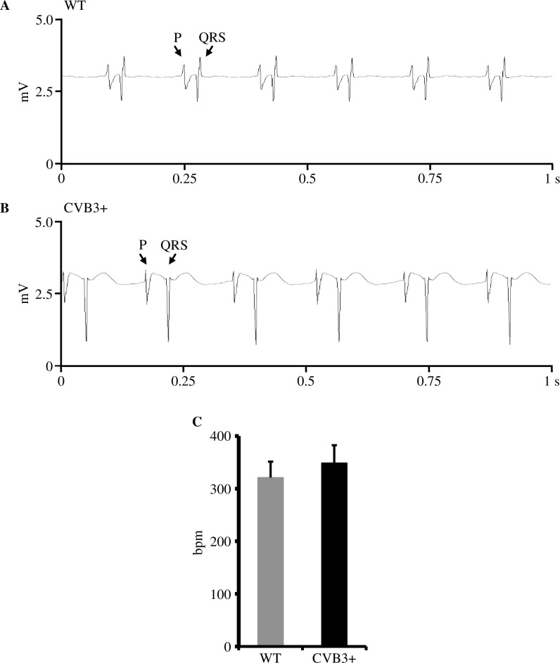
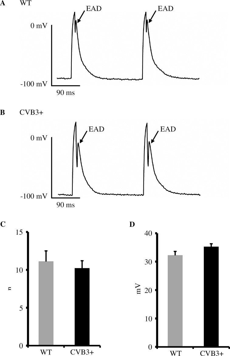
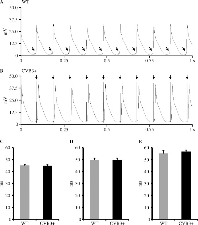
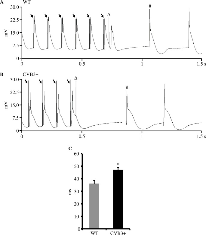
Cellular action potential duration (APD) in WT and CVB3+ myocytes was unchanged. No difference in mean occurrence or amplitude of afterdepolarizations in WT and CVB3+ myocytes was found. Interestingly, resting membrane potential in CVB3+ myocytes was significantly hyperpolarized (WT: -90.0±2.2 mV, n = 7; CVB3+: -114.1±3.0 mV, n = 14; p<0.005). Consistently, in Langendorff-perfused hearts, APDs were also not different between WT and CVB3+ whole hearts. Within both groups, we found a heart rate dependent shortening of ADP90 with increasing heart rate in Langendorff-perfused hearts. VERP was significantly prolonged in CVB3+ hearts compared to WT (WT: 36.0±2.7 ms, n = 5; CVB3+: 47.0±2.0 ms, n = 7; p = 0.018). Resting heart rate (HR) in Langendorff-perfused hearts was not significantly different between both genotypes. Electrical pacing protocols induced no VA in WT and CVB3+ hearts.
In CVB3+ mice, prolonged ventricular refractoriness and hyperpolarized resting membrane potentials in presence of unchanged APD were observed, suggesting that low level CVB3 expression does not promote VA by altered cardiac electrophysiology in this type of chronic myocarditis. These findings may suggest that other mechanisms such as chronic myocardial inflammation or fibrosis may account for arrhythmias observed in patients with chronic enteroviral myocarditis.
已知柯萨奇病毒B3(CVB3)可诱发急性和慢性心肌炎。大多数感染在临床上并无明显症状,但有些患者会出现室性心律失常(VA)和心源性猝死(SCD)。研究表明,急性CVB3感染可能导致心脏离子通道功能受损,从而形成促心律失常基质。然而,心肌细胞中低水平的CVB3+表达是否会导致心脏电生理改变进而引发VA尚不清楚。
利用细胞电生理学分析野生型(WT)和转基因CVB3ΔVP0(CVB3+)小鼠分离的心肌细胞的细胞动作电位(AP)和后去极化的发生情况。此外,我们研究了Langendorff灌注全心脏的体表心电图、单相动作电位、心室有效不应期(VERP)和VA的诱发性。所有使用的心肌细胞和全心脏均来自雄性小鼠。
WT和CVB3+心肌细胞的细胞动作电位持续时间(APD)未发生变化。WT和CVB3+心肌细胞后去极化的平均发生率或幅度没有差异。有趣的是,CVB3+心肌细胞的静息膜电位显著超极化(WT:-90.0±2.2 mV,n = 7;CVB3+:-114.1±3.0 mV,n = 14;p<0.005)。同样,在Langendorff灌注心脏中,WT和CVB3+全心脏的APD也没有差异。在两组中,我们发现Langendorff灌注心脏中ADP90随心率增加而呈心率依赖性缩短。与WT相比,CVB3+心脏的VERP显著延长(WT:36.0±2.7 ms,n = 5;CVB3+:47.0±2.0 ms,n = 7;p = 0.018)。Langendorff灌注心脏中两种基因型的静息心率(HR)无显著差异。电起搏方案在WT和CVB3+心脏中均未诱发VA。
在CVB3+小鼠中,观察到心室不应期延长和静息膜电位超极化,而APD未改变,这表明在这种类型的慢性心肌炎中,低水平的CVB3表达不会通过改变心脏电生理来促进VA。这些发现可能表明,其他机制,如慢性心肌炎症或纤维化,可能是慢性肠道病毒心肌炎患者心律失常的原因。