Yan Peng-Fei, Yan Ling, Hu Ting-Ting, Xiao Dong-Dong, Zhang Zhen, Zhao Hong-Yang, Feng Jun
Department of Neurosurgery, Union Hospital, Tongji Medical College, Huazhong University of Science and Technology, Wuhan, Hubei, China.
Faculty of Medicine, University of British Columbia, Vancouver, British Columbia, Canada; Department of Computer Science, University of Northern BC, Prince George, British Columbia, Canada.
Transl Oncol. 2017 Aug;10(4):570-577. doi: 10.1016/j.tranon.2017.04.006. Epub 2017 Jun 24.
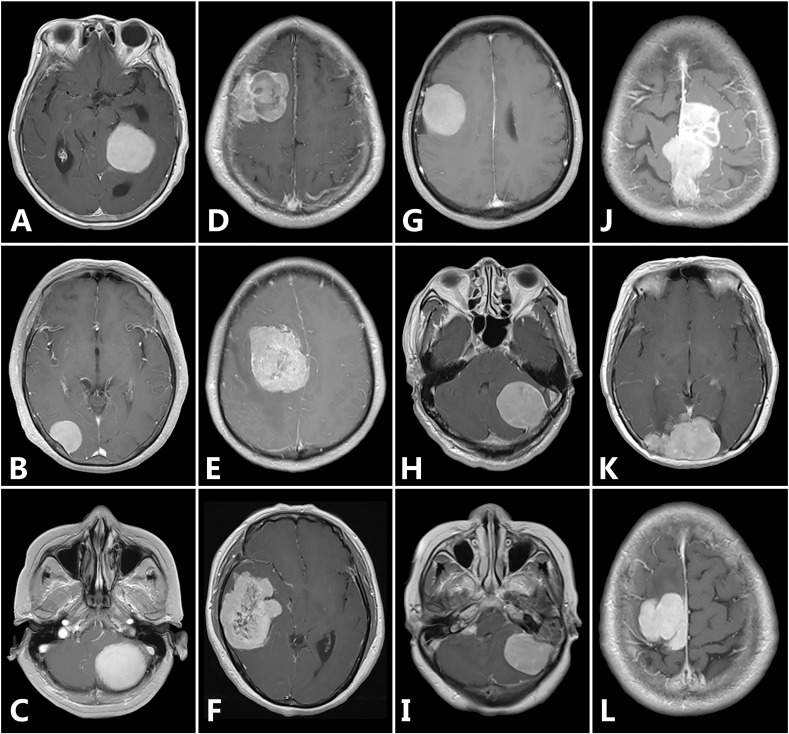
Preoperative knowledge of meningioma grade is essential for planning treatment and surgery. The purpose of this study was to investigate the diagnostic value of MRI texture and shape analysis in grading meningiomas.
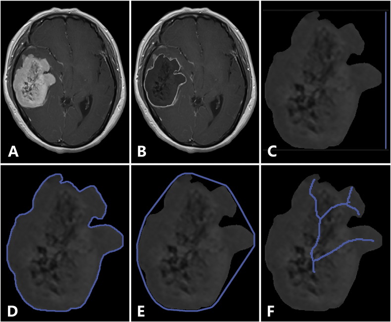
A surgical database was reviewed to identify meningioma patients who had undergone tumor resection between January 2015 and December 2016. Preoperative MR images were retrieved and analyzed. Texture and shape analysis was conducted to quantitatively evaluate tumor heterogeneity and morphology. Three machine learning classifiers were trained with these features to build classification models. The performance of the features and classification models was assessed.
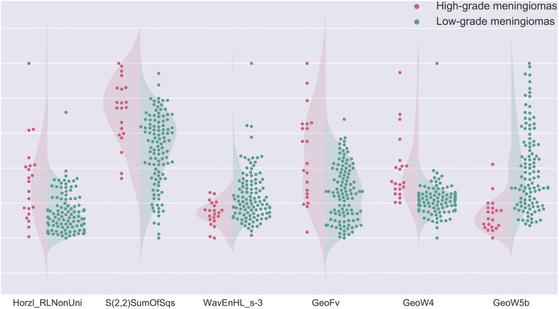
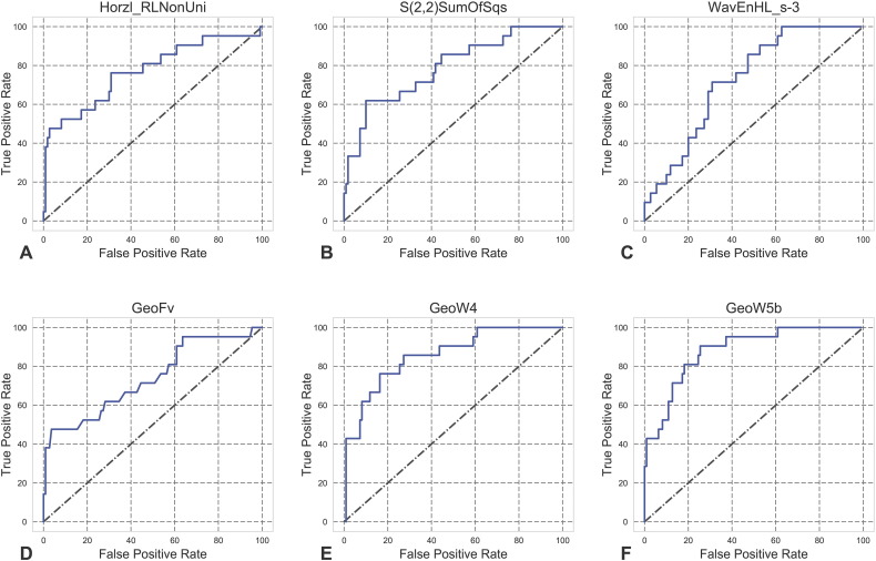
A total of 131 patients were included in this study: 21 with high-grade meningiomas and 110 with low-grade meningiomas. Three texture features were selected: Horzl_RLNonUni, S(2,2)SumOfSqs, and WavEnHL_s-3; three shape features were selected: GeoFv, GeoW4, and GeoW5b. The Mann-Whitney test indicated that all six features were significantly different between high-grade and low-grade meningiomas. AUC values were generally greater than 0.50 (range, 0.73 to 0.88). Sensitivities and specificities ranged from 47.62% to 90.48% and 69.09% to 96.36%, respectively. Among the nine classification models obtained, the one built by training the SVM classifier with all six features achieved the best performance, with a sensitivity, specificity, diagnostic accuracy, and AUC of 0.86, 0.87, 0.87, and 0.87, respectively.
Texture and shape analysis, especially when combined with a SVM classifier, can provide satisfactory performance in the preoperative determination of meningioma grade and is thus potentially useful for clinical application.
脑膜瘤分级的术前了解对于治疗和手术规划至关重要。本研究的目的是探讨MRI纹理和形状分析在脑膜瘤分级中的诊断价值。
回顾手术数据库,以确定2015年1月至2016年12月期间接受肿瘤切除的脑膜瘤患者。检索并分析术前MR图像。进行纹理和形状分析以定量评估肿瘤的异质性和形态。使用这些特征训练三个机器学习分类器以建立分类模型。评估特征和分类模型的性能。
本研究共纳入131例患者:21例为高级别脑膜瘤,110例为低级别脑膜瘤。选择了三个纹理特征:Horzl_RLNonUni、S(2,2)SumOfSqs和WavEnHL_s-3;选择了三个形状特征:GeoFv、GeoW4和GeoW5b。Mann-Whitney检验表明,高级别和低级别脑膜瘤之间的所有六个特征均存在显著差异。AUC值一般大于0.50(范围为0.73至0.88)。敏感性和特异性分别为47.62%至90.48%和69.09%至96.36%。在所获得的九个分类模型中,使用所有六个特征训练支持向量机(SVM)分类器构建的模型性能最佳,敏感性、特异性、诊断准确性和AUC分别为0.86、0.87、0.87和0.87。
纹理和形状分析,特别是与SVM分类器结合时,在脑膜瘤分级的术前判定中可提供令人满意的性能,因此在临床应用中可能具有潜在用途。