Sharma Balram, Singh Hema, Chodhary Praveen, Saran Sanjay, Mathur Sandeep Kumar
Department of Endocrinology, SMS Medical College and Hospitals, Jaipur, Rajasthan, India.
Indian J Endocrinol Metab. 2017 Jul-Aug;21(4):535-539. doi: 10.4103/ijem.IJEM_108_17.
Type 2 diabetes mellitus (T2DM) may affect bone loss differentially in adult males and postmenopausal females. We evaluated the prevalence of osteoporosis in otherwise healthy adults with T2DM.
In a cross-sectional study, adults with T2DM, aged 50 years and above, were evaluated for bone mineral density (BMD) using dual-energy X-ray absorptiometry (DXA) scan at spine and hip. T-score of ≤-2.5 was defined as osteoporosis and score -2.49 to -1.0 as osteopenia at either site. Correlation of low BMD with demographic, clinical, and laboratory parameters including serum Vitamin D and serum testosterone (in males) was evaluated.
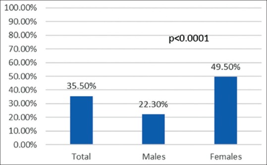
In 200 patients, mean age was 64.5 ± 7.0 years and age differed significantly in males and females ( < 0.0001). Osteoporosis was present in 35.5% adults with T2DM. Significantly greater proportion of females had osteoporosis (49.5% vs. 22.3%, < 0.0001). Frequency of osteoporosis at spine (33.5%) was higher than the same at hip (13.5%). Compared to males, significantly greater proportion of females had osteoporosis and osteopenia at both spine ( < 0.0001) and hip ( < 0.0001). Among all parameters assessed, a significant positive correlation of T-score at spine and hip was seen with body mass index in both males ( = 0.287, = 0.003 at spine and = 0.421, < 0.0001 at hip) and females ( = 0.291, = 0.004 at spine and = 0.280, = 0.010 at hip). There was no association of Vitamin D deficiency (45.5% patients) with either T-score and presence of osteoporosis either at spine ( = 0.388 and = 0.177) or hip ( = 0.431 and = 0.593).
Prevalence of osteoporosis in otherwise healthy T2DM was 35.5% with greater prevalence in females than males. Body mass but not Vitamin D or testosterone has an important role in the determination of bone loss in T2DM.
2型糖尿病(T2DM)对成年男性和绝经后女性骨质流失的影响可能存在差异。我们评估了在其他方面健康的T2DM成年患者中骨质疏松症的患病率。
在一项横断面研究中,对年龄在50岁及以上的T2DM成年患者进行脊柱和髋部的双能X线吸收测定(DXA)扫描,以评估骨密度(BMD)。任一部位T值≤ -2.5被定义为骨质疏松症,T值在-2.49至-1.0之间被定义为骨质减少。评估低骨密度与人口统计学、临床和实验室参数(包括血清维生素D和男性血清睾酮)之间的相关性。
200例患者的平均年龄为64.5±7.0岁,男性和女性的年龄差异显著(<0.0001)。35.5%的T2DM成年患者存在骨质疏松症。女性患骨质疏松症的比例显著更高(49.5%对22.3%,<0.0001)。脊柱骨质疏松症的发生率(33.5%)高于髋部(13.5%)。与男性相比,女性在脊柱(<0.0001)和髋部(<0.0001)患骨质疏松症和骨质减少的比例均显著更高。在所有评估参数中,男性(脊柱r = 0.287,P = 0.003;髋部r = 0.421,P < 0.0001)和女性(脊柱r = 0.291,P = 0.004;髋部r = 0.280,P = 0.010)的脊柱和髋部T值与体重指数均呈显著正相关。维生素D缺乏(45.5%的患者)与脊柱(r = 0.388,P = 0.177)或髋部(r = 0.431,P = 0.593)的T值及骨质疏松症的存在均无关联。
在其他方面健康的T2DM患者中,骨质疏松症的患病率为35.5%,女性患病率高于男性。体重而非维生素D或睾酮在T2DM骨质流失的决定中起重要作用。