Shin Jung-Won, Choi Hye-Ryung, Nam Kyung-Mi, Lee Hyun-Sun, Kim Sung-Ae, Joe Hyun-Jae, Kazumi Toyama, Park Kyoung-Chan
Department of Dermatology, Seoul National University Bundang Hospital, 166 Gumi-ro, Bundang-gu, Seongnam-si, Gyeonggi-do 463-707, Korea.
Department of Dermatology, Keimyung University School of Medicine, 56 Dalseong-Ro, Jung-Gu, Daegu 41931, Korea.
Int J Mol Sci. 2017 Jun 26;18(7):1360. doi: 10.3390/ijms18071360.
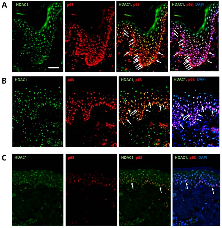
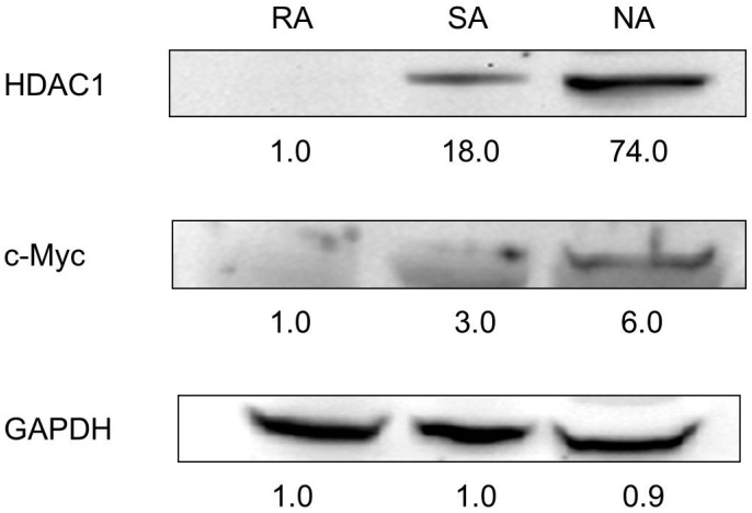
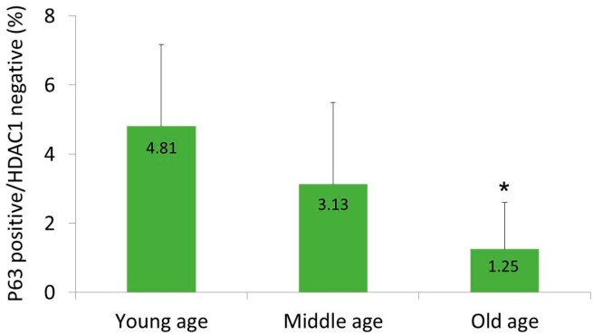
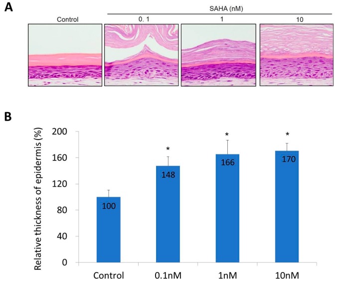
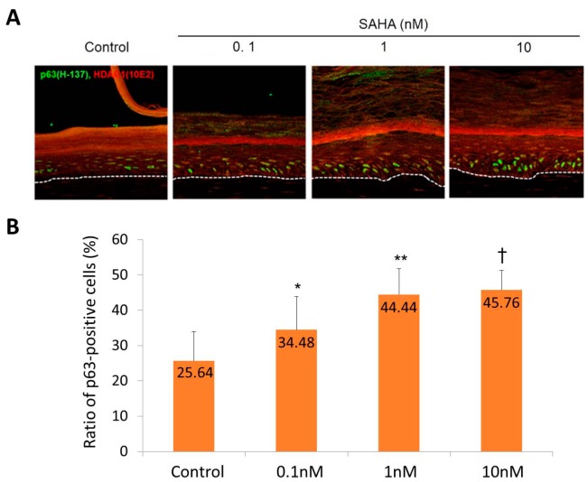
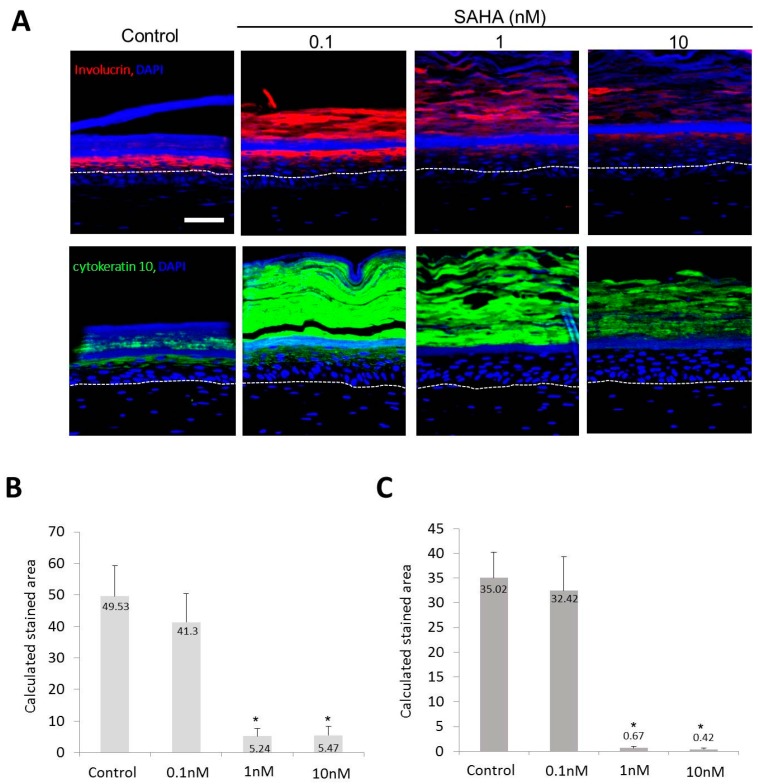
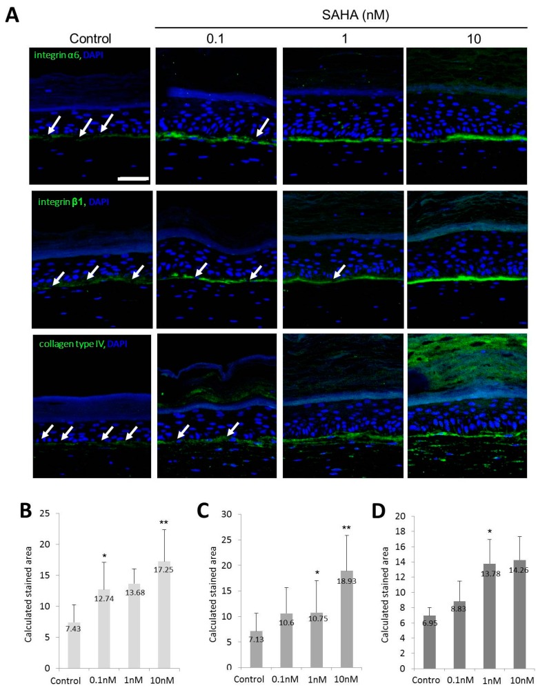
Stem cell markers of interfollicular epidermis (IEF) have not been established thus far. The aim of this study is to suggest a new way to disclose IFE-stem cells by combining the expression of histone deacetylases (HDAC) 1 and p63. Immunohistochemical staining of HDAC1 and p63 was performed in six normal human samples. Moreover, a skin equivalent (SE) model was treated with suberoylanilohydroxamic acid (SAHA, an HDAC inhibitor) to elucidate the role of HDAC1. Finally, rapidly adhering (RA) keratinocytes to a type IV collagen, which have been identified to represent epidermal stem cells, were subjected to Western blot analysis with antibodies against HDAC1. In normal samples, there was a minor subpopulation comprised of p63-positive and HDAC1-negative cells in the basal layers. The proportion of this subpopulation was decreased with age. In the SE model, SAHA treatment increased the epidermal thickness and number of p63-positive cells in a dose dependent manner. After SAHA treatment, the expression of differentiation markers was decreased, while that of basement membrane markers was increased. In a Western blot analysis, HDAC1 was not expressed in RA cells. In conclusion, the combination of p63-positive and HDAC1-negative expressions can be a potential new way for distinguishing epidermal stem cells.
迄今为止,尚未确定毛囊间表皮(IEF)的干细胞标志物。本研究的目的是提出一种通过结合组蛋白去乙酰化酶(HDAC)1和p63的表达来揭示IEF干细胞的新方法。对六个正常人体样本进行了HDAC1和p63的免疫组织化学染色。此外,用辛二酰苯胺异羟肟酸(SAHA,一种HDAC抑制剂)处理皮肤等效物(SE)模型,以阐明HDAC1的作用。最后,对已被确定代表表皮干细胞的、能快速黏附于IV型胶原的(RA)角质形成细胞,用抗HDAC1抗体进行蛋白质印迹分析。在正常样本中,基底层有一个由p63阳性和HDAC1阴性细胞组成的较小亚群。该亚群的比例随年龄增长而降低。在SE模型中,SAHA处理以剂量依赖方式增加了表皮厚度和p63阳性细胞的数量。SAHA处理后,分化标志物的表达降低,而基底膜标志物的表达增加。在蛋白质印迹分析中,RA细胞中未表达HDAC1。总之,p63阳性和HDAC1阴性表达的组合可能是区分表皮干细胞的一种潜在新方法。